Drugs, Health Technologies, Health Systems
Peer-Reviewed Article
A Review of Genomic Data Holdings in the Atlantic Provinces: Supplemental Material
Appendix 1: Inclusion and Exclusion Criteria
Please note that this appendix has not been copy edited.
Table 1: Inclusion and Exclusion Citeria
Eligibility | Inclusion | Exclusion |
|---|---|---|
Criteria |
|
|
Appendix 2: Extracted Literature on Genomic Data Collection and Analysis in Atlantic Canada
Please note that this appendix has not been copy edited.
Table 2: Genomic Datasets Collected and Analyzed in Multiple Atlantic Provinces
Article | Ethics Approval | Population and sample size | Type of sample | Test performed | Linkability/Library/Availability | Contact |
|---|---|---|---|---|---|---|
Spinal Muscular Atrophy | ||||||
McKee-Muir, Dyack, Taillon, et al. (2023)1 | IWK REB and Horizon Health Network REB. Waiver of full patient consent was obtained. | 30 patients born from 2000–2020 with SMA due to positive SMN1 from the IWK Clinical Genomics Laboratory database and MMGS. (patients from NS, NB, and PEI) | DNA: Not specified. Additional data from health records at IWK, NS SHARE, and Horizon Health Network (NB). | Performed to detect the homozygous deletion of SMN1 gene exons 7 and 8. | DNA: MMGS clinical database Heath records: Nova Scotia Secure Health Access Record (SHARE), and Horizon Health Network No statement on data storage or availability. | Jordan Sheriko, Department of Pediatrics, Dalhousie University, Halifax jordan.sheriko@iwk.nshealth.ca |
The Atlantic Partnership for Tomorrow’s Health (Atlantic PATH) study | ||||||
Sweeney, Cui, DeClercq, et al. (2017)2 | Participants were recruited between 2009 and 2015 with REB approvals in place for all 4 Atlantic provinces. Participants provided consent to be followed over a 30-year period and use of their data by Canadian and international researchers, and a research platform was designed specifically for this purpose. | Harmonized dataset for Atlantic PATH includes 34,000+ participants aged 30 to 69 (expanded to 74 after 2017). |
| Once processed, plasma, buffy coat, red blood cells, serum, saliva and urine were transferred to 2-ml cryovials and stored in −80°C freezers. Toenail samples were collected in Ziplock bags and stored in cabinets at room temperature *Genotyped data are currently available for 1000 participants. | Can be linked to health data from administrative health databases:
| Application for data process is found here: Data Access Process – Atlantic PATH |
Table 3: Genomic Datasets Collected and Analyzed in NS
Article | Ethics Approval | Population and sample size | Type of sample | Test performed | Linkability/Library/Availability | Contact |
|---|---|---|---|---|---|---|
Lynch Syndrome/Endometrial Cancer | ||||||
Levesque, Wood, Carter, et al. (2023)3 | Ethics approval from the Nova Scotia Health Research Ethics Board (1026033). *Consent not noted. | Nova Scotians diagnosed with endometrial cancer between 2017 and 2020. Tumour IHC staining was performed in 444 patients. 98 Received germline testing, 9 with germline tested had Lynch syndrome mutation | DNA sample (germline testing): Not stated Tumor biopsy and staining Also: Age, body mass index, tumour mismatch repair protein immunohistochemistry results, personal and family histories (from patient charts and Tupper gynecologic database (internal database) | Specific genetic tests and level of availability of data to researchers not stated. Germline testing results, and personal and family histories of Lynch-associated malignant disease for all eligible patients were extracted from medical genetics records by an endometrial cancer, MMGS genetic counsellor. Data used in this study are available in an unidentified, coded format from the first author on reasonable request: Marianne.levesque@dal.ca. | Dr. Katharina Kieser Dalhousie University; Maritime Medical Genetics Service, IWK Health Centre; Division of Gynaecologic Oncology, Department of Obstetrics and Gynaecology, Dalhousie University, Halifax, NS kkieser@dal.ca | |
Fetal structural anomalies via Ultrasound | ||||||
Allen, Schollenberg, Aberg, and Brock (2025)4 | IWK Health Research Ethics Board approval obtained May 21, 2019 (File #1024606), with annual renewal approval. IWK Health REB approved the Waiver of Consent due to impracticability. | Retrospective cohort (derived from the IWK Health Clinical Genomics Laboratory Information System) of 593 fetuses that had genetic testing performed due to structural anomalies identified by ultrasound in the Maritime Provinces from 2014 to 2022. | DNA Samples: chorionic villus sampling, amniocentesis, fetal blood, and products of conception Data combined with maternal health records review, EMR chart reviewed when appropriate. | First tier testing included rapid aneuploidy detection (RAD) and CMA, with subsequent sequence-based testing reserved for those cases meeting the selected testing criteria. Results of genetic testing, including RAD, karyotype, microarray (done in-house at IWK Health CGL), and gene sequencing (Single genes and small gene panels performed at the IWK CGL, larger gene panels and exomes performed and reported by referral laboratories.) For each eligible prenatal, cord, and newborn sample, an electronic health records chart review identified specific ultrasound findings, findings from other imaging modalities, involvement with medical genetic specialties, postnatal findings, and clinical outcomes including gestational age at delivery and pregnancy complications placental insufficiency, as well as newborn examination and autopsy findings. Dr. Victoria Allen Department of Obstetrics and Gynaecology Dalhousie University and IWK Health, Halifax, Canada victoria.allen@dal.ca | Dr. Victoria Allen Department of Obstetrics and Gynaecology Dalhousie University and IWK Health, Halifax, Canada victoria.allen@dal.ca | |
Meier-Gorlin syndrome | ||||||
Guernsey, Matsuoka, Jiang, et al., (2011)5 *Letter | Approval for the research study was obtained from the IWK Health Centre and CHU Ste-Justine research ethics boards. Informed consent was obtained for all participants in the study. | 5 individuals with Meier-Gorlin syndrome: 1 Maritime-Acadian, 2 Quebec and 2 Louisiana-Acadian families (individuals 1652, 1768, 1882, 1899 and 1627) 314 local Maritime control chromosomes, which (include a substantial proportion of Acadians). | DNA Sample: blood samples Also, anthropometric measurements | High density genome-wide SNP genotyping using Illumina Human 610K panel of approximately 600,000 markers, used PLINK to search for shared haplotypes among subsets of the genotyped individuals Case and control samples sequenced at McGill University and Genome Quebec Centre for Innovation. Sequence traces analyzed using Mutation Surveyor. | Illumina used to sequence data. Data from this study deposited in the RefSeq database under the following accession codes: human ORC4 mRNA, NM_002552; human ORC4 protein, NP_ 002543; human ORC1 mRNA, NM_004153.3; human ORC1 protein, NP_004144.2; human CDT1 mRNA, NM_030928.3; human CDT1 protein, NP_112190.2. | Mark E Samuels mark.e.samuels@umontreal.ca |
Renal cell carcinoma (RCC) | ||||||
Kokorovic, Thomas, Serrano-Lomelin et al., (2020)6 | No research ethics approval listed. *Secondary data sources, no patient consent. | 75 patients/family members who were evaluated for hereditary RCC syndrome and received genetic testing from January 2006 until December 2017 (collected retrospectively) received genetic testing | Clinical, pathological, and genetic analysis data. | Panel for genetic testing including FH, FLCN, MET, PTEN, SDHB, SDHC, SDHD, TMEM127, and VHL genes. Genetic testing was performed by the Clinical Genomic Laboratory, IWK, Halifax; Leeds Genetics Laboratory (Molecular Genetics), St. James’ University Hospital, Leeds, UK, or Blueprint Genetics, Helsinki, Finland. | *Level of detail with genetic results available to researchers unclear *No statement on data availability. | Dr. Ricardo A. Rendon Queen Elizabeth II Health Sciences Centre, Halifax, NS, Canada; rrendon@dal.ca |
Merkel cell carcinoma | ||||||
Walsh (2001)7 Ly, Walsh, and Pasternak (2012)8 Fleming, Ly, Pasternak et al. (2014)9 Carter, Gaston, Huang, et al. (2018)10 DeCoste, Carter, Pasternak, et al. (2021)11 DeCoste, Walsh, Gaston, et al., (2022)12 | study approved by the Capital Health Research Ethics Board, Halifax, Nova Scotia. (REB #1021594) *No mention of participant consent to study or secondary use. | Total of 142 study cases submitted through the Pathology Department of the Victoria General Hospital/Queen Elizabeth II Health Sciences Centre in Halifax, identified through a search of laboratory computer records and review of a previously published case series of MCCs. All cases diagnosed between 1989 and 2020. Only those with available paraffin-embedded tissue samples included. 42 primary MCCs,13 MCC metastases, 57 non-MCC cutaneous lesions, 15 pulmonary neuroendocrine carcinomas and 15 GI NETs were included. | Clinical information was obtained from pathology requisition forms, requesting physicians, and pathology reports. Thirteen MCC metastases were examined. Ten of these metastases were derived from 8 primary PMCC and 2 primary CMCC tumours already included in the study. | No genetic testing conducted. IHC was performed on paraffin-embedded archival tissue to detect positivity for CK20 and CM2B47 For 83 cases in 2014, new slides were cut from archived paraffin-embedded tissue blocks and stained with p63 as per the manufacturer's instructions (Clone BC4A4 [Biocare Medical, Concord, CA), CC1 antigen retrieval and iView Kit detection method [Ventana])9 46 cases, 21 pure Merkel cell polyomavirus (MCPyV)+, 10 pure MCPyV−, and 15 combined MCPyV− MCCs. DNA (80 ng) from each sample was probed with SNP-containing oligonucleotides targeting interrogation sites of 40 base pairs. Genome-wide CNA studies were performed for from 28 MCCs (9 MCPyV+, 9 MCPyV−, and 10 combined tumours). DNA from all 46 cases was analyzed by NGS using a custom set of primers for 8 tumour suppressor genes and 1 oncogene previously shown to have inactivating and activating mutations in MCC, (TP53, RB1, APC, NOTCH1, NOTCH2, MLL2, MLL3, BRCA1,and PIK3CA)10 NGS of 51 cases were selected primary cutaneous Merkel cell carcinomas (21 MCPyV +, 13 pure MCPyV− and 17 combined MCPyV−) derived from the Maritime region of Canada (1993 to 2020).extracted DNA was analyzed via the Illumina TSO500 hybrid capture DNA panel, performed on an Illumina NextSeq550 instrument (San Diego, CA), in accordance with the manufacturer's instructions. The panel analyzes 523 cancer-related genes for small mutations, a subset of genes for copy number gains, as well as global parameters of tumour mutation burden (TMB) and microsatellite instability (MSI) Cases with median exon coverage < 100x (unique reads employed for all thresholds) were excluded from analysis11,12 | *These cases were obtained through medical points of contact and patients required information on tests performed, therefore these are likely likable to administrative data. DNA was extracted using QIAamp DNA FFPE (Qiagen Inc, Valencia, CA) or MagNA Pure Compact (Roche, Basel, Switzerland) commercial kits and quantitated using the Qubit dsDNA BR kit (Thermo Fisher Scientific Inc, Grand Island, NY). Affymetrix (Santa Clara, CA), conducted genome-wide CNA studies using the OncoScanFFPE gene chip probe array, Next-generation DNA sequencing data were processed using an in-house bioinformatics analysis pipeline conducted on an Illumina MiSeq gene sequencer (San Diego, CA). Processed using Picard and the Genome Analysis Toolkit following community-developed best practices. Copy number aberration data from each sample were analyzed using Nexus Copy Number version 9.0 (BioDiscovery, El Segundo, CA).10 Data were first analyzed using Illumina's TSO500 LocalApp workflow for alignment and variant calling of mutations 33 cases (9 MCPyV+, 10 pure MCPyV− and 14 combined MCPyV−) were sent to Mayo Clinic Laboratories (Rochester, MN) for Rb immunohistochemical (IHC) staining, which was performed in accordance with their institutional protocols. The datasets used and/or analyzed during the current study are available from the corresponding author on reasonable request. 11,12 | Dr. Sylvia Pasternak Dalhousie University Department of Pathology Halifax, Nova Scotia, sylvia.pasternak@nshealth.ca Noreen M.G. Walsh, MD, Division of Anatomical Pathology, Queen Elizabeth II Health Sciences Centre, Halifax, NS Canada Dr. Ryan C. DeCoste Department of Pathology and Laboratory Medicine, QEII Health Sciences Centre), Halifax, NS, ryan.decoste@dal.ca |
Exfoliation syndrome | ||||||
Li, Wang, Lee et al., (2021)13 | Patients with exfoliation syndrome were enrolled following informed consent. Both studies approved by the REB of the NSHA (protocols 1011873 and 1020512). | Multiple cohorts from multiple countries included 403 cases, 392 controls included from NS (Supplemental materials) Reference controls were obtained from the DNA diagnostic laboratory serving the same population. All participants were of self-reported European ancestry. | Venous blood samples Eye testing results | NS participants underwent whole exome Sequencing during first validation stage | Whole-exome sequencing and targeted sequencing (targeting the CYP39A1 [Refseq NM_016593] coding sequence) libraries were prepared using hybridization capture kits (Roche-Nimblegen SeqCap) Cases and controls sequenced together using 2 × 151 base-pair, paired-end reads on Illumina instruments. Raw DNA sequence reads from the Illumina HiSeq 4000 and Novaseq 6000 instruments were aligned to the hg19 genome build. Variant detection was performed using exons and 50 base-pair regions flanking the exons. DNA sequence reads were mapped using Burrows-Wheeler Aligner software (version 0.7.16a-r1181). Variant calling was performed using the Genome Analysis Tool Kit (version 3.7) and the Picard (v *No statement of data availability *unclear if data can be linked to administrative data | Chiea Chuen Khor, MD, Ph.D., Genome Institute of Singapore, 60 Biopolis St, 02-01 Genome Bldg, Singapore 138672 (khorcc@gis.a-star.edu.sg |
Aung, Ozaki, Lee et al., (2017)14 | For Canada, individuals with Exfoliation syndrome were enrolled following informed consent from within the Nova Scotia Health Authority (NSHA), Nova Scotia (protocols 1011873 and 1020512) | World-wide GWAS partnership study (Canada): 341 cases and 247 controls Canada Replication:136 cases, 747 controls All participants were of self-reported European ancestry | DNA samples obtained from ocular tissues | Deep sequencing was performed on a total of 5,570 XFS cases and 6,279 controls from 9 countries (excluding Canada (NS)). A genome-wide association study (GWAS) of Exfoliation syndrome cases and controls from 24 countries (including Canada) followed by replication in 18 countries (including Canada) identified 7 genome-wide significant loci. | Genome-wide genotyping was undertaken using the Illumina OmniExpress BeadChip. All 9,035 XFS cases and 17,008 controls for the GWAS discovery stage genotyped using Illumina OmniExpress array. In the replication stage, direct genotyping using the Sequenom Mass-Array and Applied Biosystems Taqman genotyping platforms was carried out for cases and controls from Nova Scotia (Supplemental materials). | Tin Aung Dept of Ophthalmology, Yong Loo Lin School of Medicine, National University of Singapore, Singapore aung.tin@singhealth.com.sg Francesca Pasutto Institute of Human Genetics, Friedrich-Alexander-Universität, Erlangen-Nürnberg, Erlangen, Germany Francesca.Pasutto@uk-erlangen.de Janey L. Wiggs Department of Ophthalmology, Harvard Medical School, Massachusetts Eye and Ear Infirmary, Boston, Massachusetts, US Janey_Wiggs@meei.harvard.edu Chiea Chuen Khor Genome Institute of Singapore, Singapore khorcc@gis.a-star.edu.sg |
Multiple myeloma (MM) | ||||||
Cutler, Knopf, Campbell, et al., (2021)15 | Conducted under ethical approval by the Nova Scotia Health Authority (NSHA) Research Ethics Board (1021520 and 1021397), and patients provided written informed consent for research. | 77 patient samples (from 76 patients) bone marrow was collected from patients with plasma cell dyscrasias at the Victoria General Hospital (Halifax, NS, Canada) | Patient bone marrow samples laboratory data, including albumin, Beta2-microglobulin, lactate dehydrogenase, bone marrow plasma cell burden, serum M-protein quantity, immunoglobulin heavy and light chain type and quantity, serum free light chain ratio, and FISH data, coinciding with the time of bone marrow acquisition, were collected from the NSHA laboratory information system. Patient clinical data, including age, sex, diagnosis at time of bone marrow acquisition, therapies received, follow-up period, stage, and time of events, were also collected from the NSHA hospital information system. | A custom panel to target exons of these 26 genes was designed using Illumina DesignStudio (Illumina, San Diego, CA).genes that were mutated in > 1% of the population of patients were included (25 genes) in published WES and WGS studies as well as MYC. | MM-specific targeted-sequencing approach using a custom-designed 26-gene panel, the DMG26, that is applicable in a standard clinical molecular laboratory and demonstrates that the DMG26 captures prognostically relevant genomic abnormalities that are distinct from those captured by FISH. Genomic data generated during this study are available on request, with access managed by the Dalhousie Pathology Biobank. All programming scripts used in this work are available at GitLab, last accessed November 25, 2020). DNA library preparations for the DMG26 panel were performed per Illumina TruSeq and AmpliSeq for Illumina custom panel reference guides. Libraries were sequenced in 2 runs on an Illumina MiSeq at a mean depth of approximately 1000 times. FastQ files were analyzed using an in-house bioinformatic pipeline Data is linkable to administrative health data | Daniel Gaston, Ph.D., 5788 University Ave., Halifax, NS, Canada. dan. gaston@nshealth.ca |
Familial exudative vitreoretinopathy (FEVR) | ||||||
Robitaille, MacDonald, Kaykas, et al., (2002)16 | IWK Health Centre Research Ethics Board and the Oakland University Institutional Review Board of Human Subjects Investigation Committee approved these studies. Informed consent obtained. | 81 subjects: 27 people with FEVR from NS and 54 of their family members without FEVR | DNA from blood samples | Determined genotypes of 8 family members using 6 DNA markers (D11S1887, D11S896, D11S4082, D11S1780, D11S873 and D11S1311) from the EVR1 locus. Performed Linkage analysis after collection of samples from additional family members with 4 Genethon (D11S1887, D11S4082, D11S1780, D11S1311) and 2 Marshfield markers (D11S896, D11S873). Examined an additional 11 Genethon, 7 CHLC and 9 novel CA-repeat markers (33 total)(fine mapping). Haplotype construction using 33 polymorphic markers revealed 5 proximal and 4 distal recombination’s (5 were affected, 4 unaffected). Analyzed the coding regions of FZD4 and FLJ22104 for mutations 3 family members (2 affected, 1 unaffected) by sequencing PCR amplified fragments. Used PCR–RFLP analysis and fluorescence capillary electrophoresis to screen the remainder of the family and a control population for presence of mutations observed in the affected individuals. Detected both the M493_W494del and L501fsX533 mutations. | Genotyping using marker sequences from the Genome Database on an Applied Biosystems Prism 3100 Genetic Analyzer running GeneMapper software (Applied Biosystems), and we verified mendelian inheritance of alleles using the PedCheck program performed two-point linkage analysis using the MLINK program version 4.1p from the FASTLINK software package, and multipoint linkage analysis using VITESSE version 1.0 and SIMWALK version 2.8, assuming a disease allele frequency of 0.000 Designed intronic primer pairs for each exon in FZD4 and FLJ22104 using the Primer3 program. Used an Applied Biosystems Prism 3100 Genetic Analyzer to detect M493_W494del and L501fsX533 mutations. | Dr. Michael R. Hayden Xenon Genetics, Inc., Burnaby, BC mrh@cmmt.ubc.ca |
Ells, Guernsey, Wallace et al., (2010)17 | Approvals obtained from IWK Health Center, Calgary Health Region (Alberta Children’s Hospital site), and the St Joseph Health Care London. Consent obtained according to the Canadian Tri-Council guidelines. | 104 premature infants recruited both prospectively and retrospectively between March 1, 2003 and April 30, 2008, 71 with severe ROP, 33 mild to no ROP. 173 random Canadian samples *Number from NS not stated | Tissue samples (blood or buccal swabs) | Direct automated sequencing of the 2 coding exons of the FZD4 gene. Mutation detection was performed both manually and using Mutation Surveyor | Primers can be requested from the corresponding author. *No other statements of data availability. | Johane Robitaille Eye Care Team, IWK Health Center, University Ave. Halifax, Nova Scotia Canada. E-mail: jrobitai@dal.ca |
Robitaille, Gillett, LeBlanc et al., (2014)18 | Approved by IWK REB. Written informed consent obtained from participant | 72 probands recruited from April 1998-September 2013. Unclear how many from Halifax. | DNA extracted from blood or saliva samples Collected clinical data retrospectively. | WES conducted. Also direct automated Sanger sequencing of 2 coding exons of the FZD4 gene, the 23 coding exons of the LRP5 gene, and 22 coding exons of the KIF11 gene. | WES conducted at McGill University and Genome Quebec Innovation Centre. DNA sequenced with 100 base pair paired-end reads (HiSeq 2000 sequencer; Illumina). Genomic variants called using Genome Analysis Toolkit pipeline and annotated with ANNOVAR. Specific exons sequenced using Sanger sequencing performed with ABI PRISM 3100 Genetic Analyzer and mutation calling was done by Mutation Surveyor, version 3.97) | Dr. Johane M. Robitaille University Avenue, PO Box 9700, Halifax, NS, jrobitai@dal.ca. |
van der Ende, Bedard, Wallace, et al., (2025)19 | Approved by the REB of the IWK Health Centre. Consent obtained in accordance with Declaration of Helsinki. | Patients with retinal features compatible with FEVR, affected relatives, and 1st-degree relatives were recruited between 1998 and 2024. Excluded people with retinopathy of prematurity. 94 people (76 had WES, 18 had Sanger sequencing data.) | DNA was extracted from peripheral blood or saliva samples Also: results from eye examinations and IV fluorescein angiography and fundus photography (if available). Ethnicity provided at discretion of referring physician. | Sanger sequencing was used to identify rare variants in known genes, and this method alone identified a molecular diagnosis for 18 probands. 76, including 2 diagnosed with Sanger sequencing in prior publications. A virtual FEVR gene panel was used in those 76 probands to identify variants in FZD4, LRP5, TSPAN12, NDP, ZNF408, CTNNB1, KIF11, ATOH7, ILK, JAG1, TGFBR2, RCBTB1, DOCK6, ARHGAP31 (, NOTCH1, CTNNA1, CTNND1, DLG1, COL9A1, LRP6, TUBGCP4, TUBGCP6, PLK4, CDK19, and LAMA1. At minimum, the Gene, Nucleotide Change, Amino Acid Change, Variant Type, REVEL, CADD, SpliceAI, gnomAD, Sex, ACMG ranking, SVI evidence were collected. | WES completed using Illumina NextSeq 550. Genomic variants called using the Genome Analysis Toolkit and Picard. Variants were annotated with Annovar and compared against dbSNP, 1000 Genomes project, and the genome aggregation dataset version 4. No statement of data availability. Data likely linkable to administrative health data as patents recruited through health care system. | Dr. Johane M. Robitaille University Avenue, PO Box 9700, Halifax, NS, jrobitai@dal.ca. Christopher R. McMaster, Department of Pharmacology, Dalhousie University Halifax, NS, christopher.mcmaster@dal.ca |
Colonic mixed adenoneuroendocrine carcinoma and neuroendocrine carcinoma | ||||||
Sinha, Gaston, Manders, et al., (2018)20 | Approved by our institutional research ethics board. | All colonic tumours (269?) with a previous diagnosis of MANEC, high-grade NEC, and adenocarcinoma with neuroendocrine differentiation were retrieved from the archives of the Department of Pathology, QE II Health Sciences Center, between July 2000 and July 2015. 269 colorectal adenocarcinomas used as controls | Tumor samples. Samples deidentified, and tissue cores obtained from an area containing at least 80% viable tumour cells. Immunohistochemistry (IHC) of tumour sections | Sought to identify unique chromosomal alterations of MANEC and NEC by comparing their genome-wide CNAs to those of 269 conventional adenocarcinomas characterized the genome-wide copy number aberrations of 14 MANECs and 5 neuroendocrine carcinomas using the OncoScan FFPE (Affymetrix, Santa Clara, CA) assay. Genomic Identification of Significant Targets in Cancers algorithm identified 7 peaks that drive the tumorgenesis of MANEC. Genomic Identification of Significant Targets in Cancer (GISTIC) analysis was used to distinguish significant chromosomal aberrations from random background. CNA data generated for colonic MANEC and NEC were compared colorectal adenocarcinomas. For statistical comparison of copy number alternations between MANEC and adenocarcinoma, false discovery rate–corrected Ps were then obtained using the Benjamini-Hochberg procedure. MSI kit from Promega (Madison, WI): The presence of the BRAF V600 mutation was detected using a SNaPshot-based assay that can detect the V600E, V600K, V600M, V600R, and V600D mutations | comparison colorectal adenocarcinomas investigated and analyzed by the OncoScan FFPE assay and Nexus copy number software the OncoScan FFPE assay from Affymetrix (Santa Clara, CA) was performed on 19 samples. Raw data analyzed using Nexus Copy Number 7.5 Discovery Edition software (BioDiscovery, El Segundo, CA). | Dr. Weei-Yuarn Huang Division of Anatomical Pathology, Queen Elizabeth II Health Sciences Centre and Dalhousie University, Halifax, Nova Scotia, Canada B3H 1V8. huangw@nshealth.ca |
Bipolar disorder | ||||||
Hou, Heilbronner, Degenhard, et al. (2016)21 Stone, Nunes, Akiyama, et al. (2021)22 O’Connell, Lee, Koromina, et al. (2025)23 | Written informed consent was obtained from all participants. Ethical and regulatory approvals were obtained at each site that contributed anonymized data and DNA to the analysis. | GWAS of lithium response in 2563 patients with DSM III or DSM-IV diagnosis of a bipolar spectrum disorder, collected by 22 participating sites from the International Consortium on Lithium Genetics. Patients required to have taken lithium for ≥ 6 months with no additional mood stabilizer (2008 and 2013). One site was Halifax. They conducted GWAS in 2 parts: Halifax: 353 total, GWAS 1: 241, GWAS 2: 112 | Whole blood samples. assessment methods: semi-structured clinical interviews (clinical), medical records, registries and questionnaire data (community) and self-reported surveys) | largest-ever set of genomic data from BD patients treated with lithium for maintenance therapy over a duration of at least 1 year, from 14 international site members of the Consortium on Lithium Genetics (ConLiGen) and 79 cohorts. A total of 3193 participants were genotyped; 2563 remained after quality control (1162 in GWAS 1 and 1401 in GWAS 2). Data from common single nucleotide polymorphisms (SNPs) were tested for association with categorical and continuous ratings of lithium response. Lithium response was measured using a well established scale (Alda scale). Identified 337 linkage disequilibrium-independent genome-wide significant variants that map to 298 loci | Samples were genotyped at the National Institute of Mental Health (Bethesda, MD, US), Life and Brain Center at the University of Bonn (Bonn, Germany), or Broad Institute (Cambridge, MA, US) using either Affymetrix or Illumina SNP arrays Halifax: 241 individuals genotyped with an SNP array from Illumina Omni1-Quad in GWAS1 and 112 individuals genotyped with Illumina OmniExpress 1·1 in GWAS 2 (supplementary materials). After basic quality control and linkage disequilibrium (LD) pruning, about 40K directly genotyped SNPs shared across all SNP arrays were used for the relatedness testing by PLINK. | Thomas G Schulze, Institute of Psychiatric Phenomics and Genomics (IPPG), Medical Center of the University of Munich, Munich, Germany thomas.schulze@med.uni-muenchen.de |
Youth Anxiety disorders | ||||||
McAusland, Burton, Bagnell, (2024)24 *Study protocol* Not yet conducted | Eligible youth will complete the consent form and may opt-in to long-term storage of their DNA sample. In some cases parental consent will be obtained with youth assenting to participate. | The goal is to recruit 13,000 youth aged 10 to 19 of which 50% are expected to endorse symptoms of anxiety that indicate the presence of an anxiety disorder. The GAYA study with local sites in Calgary, Halifax (IWK Health Centre community), Hamilton, and Vancouver. Clinical recruitment approach and online recruitment (Social media content and visuals) | An OG-600 Oragene saliva DNA sample kit or choose to have an OCR-100 ORA collect DNA sample kit mailed to their home with a pre-paid return envelope. Also, participants will be asked to complete the Screen for Child Anxiety Related Emotional Disorders (SCARED), the Short Mood and Feeling Questionnaire (SMFQ), the CPSS-SR-5, the Behavioural Inhibition and Behavioural Activation System (BIS/BAS) scales. Also 5 optional surveys. | Genetic Architecture of Youth Anxiety (GAYA): De-identified research IDs will link data and saliva samples. The linked IDs will be logged in REDCap to document and track the handling of biologic samples. DNA extraction from saliva is performed at each individual site following best practice and according to the manufacturers’ protocols. Subsequent genotyping will be performed in 3 batches. All individuals will be genotyped. | genotyped (using DNA from the saliva samples) with Illumina’s Global Screening Array v3.0 (GSA). For genotyping calling Illumina’s GenomeStudio will be used. Spit for Science genotyping will be done locally using the same Illumina array and their genetic data are sent to the Halifax site once the participant has completed the questionnaires and/or app portion of the study. | Dr. Laina McAusland Department of Psychiatry, University of Calgary, Calgary, AB, Canada |
Autosomal Dominant kidney disease | ||||||
Kmochová, Kidd, Orr, et al. (2024)25 | Approved by Nova Scotia Health REB | 5 families found to have autosomal dominant medullary amyloidosis due to 2 different pathogenic APOA4 variants. From sites in United States, Czech Republic, and Nova Scotia, Canada (at least 32 people from 1 family) | Whole blood or whole saliva | Whole exome of whole genome sequencing underwent whole genome sequencing with identification of a chr11:116692578 G>C (hg19) variant encoding the missense mutation p.L66V of the ApoA4 protein. | Sanger Sequencing | Dr. Anthony Bleyer Wakefeild School of Medicine Winston-Salem, North Carolina ableyer@wakehealth.edu |
Table 4: Genomic Datasets Collected and Analyzed in NB
Article | Ethics Approval | Population and sample size | Type of sample | Test performed | Linkability/Library/Availability | Contact |
|---|---|---|---|---|---|---|
Breast carcinoma | ||||||
Ouellette, Richard, and Maïcas, (2004)26 | REB approval not specified. Consent for both surgical procedures and SLN analysis was obtained from all patients. | 53 consecutive patients (42 patients retained for analysis) with breast CA (cases) and 25 patients with melanoma (controls) at CHU Dumont from 1999 to 2002. | SLN biopsy | RNA and DNA from samples assessed for genes coding for MGB1 and MGB2 using PT-PCR testing. | Not specified. | Not specified. Dr. Rodney Ouellette, MD Ph.D. Dr Georges-L. Dumont Hospital University of Moncton |
Gauvin, Allain, Bouhamdani, Williams, et al. (2025)27 | Approved by the Vitalité Health Network REB (#101703). No consent due to retrospective design | 445 participants from NB (Acadian and French Canadian, European descent (non-French or Acadian), South-American, Native American, African or Asian origins) tested for hereditary breast C gene from 2019 to 2022 | Not specified. Data on other health information collected from patient charts. | Multi-gene panel testing (268), or targeted predictive testing for a known familial variant (39) for genes known to be associated with breast CA. Genetic testing performed by Fulgent Genetics or GeneDx. | Paired-end sequencing with Illumina sequencers. Data is available upon request. | Eric Allain eric.allain@vitalitenb.ca Mouna Ben Amor mouna.benamor@vitalitenb.ca |
Myeloid neoplasms | ||||||
Northrup, Maybank, Carson, et al. (2020)28 | Approved by the Horizon Health Network REB | 178 (156 from Saint John, 22 other locations) randomly selected specimens from patients with Myeloid Neoplasms submitted for the NGS Ion AmpliSeq Myeloid Panel (2017) or the Oncomine Myeloid Panel (Thermo Fisher)(2018) | 43 bone marrow aspirates and 136 peripheral blood samples | NGS of a genetic panel of genes related to hematologic malignancies Additionally, karyotyping done in 75 cases | Library preparation performed using Ion AmpliSeq Myeloid Panel (145 samples) or Oncomine Myeloid Panel (33 samples) at the Saint John Regional Hospital. | Victoria Northrup, MSc, Dalhousie Medicine NB, Saint John, Canada; and Departments of Laboratory Medicine and Research Services, Saint John Regional Hospital, Saint John, Canada. |
Healthy participants with Acadian heritage | ||||||
Robichaud, Allain, Belbraouet et al. (2022)29 | Vitalité Health Network REB Consent obtained from all subjects for DNA collection and project participation | 60 from Southeast NB (all at least 1 Acadian grandparent born in the region) *Plans to expand to more areas of NB | DNA samples collected using the Oracollect DNA buccal swab from DNA Genotek inc. | DNA from oral swabs was extracted by Fulgent Genetics. WES of 312 autosomal recessive and 30 X-linked diseases connected with French ancestry | NGS libraries generated using modified KAPA DNA Library. Prepared DNA libraries sequenced using a HiSeqX or NovaSeq 6000 from Illumina inc. (San Diego, CA, US). Data restricted due to higher risk of re-identification from genetic information | Dr. Mouna Ben Amor Department of Medical Genetics, Vitalité Health Network, Dr. Georges L.-Dumont University Hospital Centre, Moncton mouna.benamor@vitalitenb.ca |
Pancreatic ductal adenocarcinoma | ||||||
Roy, Wajnberg, Ouellette, et al. (2023)30 | Approval obtained from Vitalité Health Network REB consent obtained from patients | 16 PDAC patients and 13 non-affected individuals | Plasma samples | Assessed utility of the Vn96 synthetic peptide for the isolation of extracellular vesicles from human plasma and the subsequent detection of small RNA biomarkers of PDAC by NGS analysis. | Tumoral Biobank at CHU Dumont and the Ontario Pancreas CA Study at Mount Sinai Hospital (Toronto, ON). No statements of data availability. | Stephen M. Lewis Atlantic Cancer Research Institute, Moncton, NB, Canada Beatrice Hunter Cancer Research Institute, Halifax, NS, Canada Department of Chemistry and Biochemistry, Université de Moncton, Moncton, NB, Canada stephenl@canceratl.ca |
Table 5: Genomic Datasets Collected and Analyzed in NL
Article | Ethics Approval | Population and sample size | Type of sample | Test performed | Linkability/Library/Availability | Contact |
|---|---|---|---|---|---|---|
General Newfoundland and Labrador population | ||||||
Zurel, Bhérer, et al. (2025)31 Gilbert, Zurel, et al. (2023)32 | NLGP protocol was approved by the NL Health REB (#: 2018.243). Participants consented to secondary use, pending access requests for scientific research through Sequence Bio board. | Random recruitment of participants from general practice clinics across the province aged ≥ 18 years, possessed a valid NL health card, and provided written consent. 2,500 participants total, including people of Indigenous heritage, 1,807 NL individuals of European descent for the NLGP cohort. Additionally, included data from the Irish DNA Atlas and the People of the British Isles. | Saliva samples were obtained and medical histories provided by all 2,500 participants. Information also collected on religion and the birthplace of their parental ancestors. | Targeted SNP genotyping: 1,110 Y chromosomes from an NL-based cohort were analyzed using 5,761 Y-specific SNPs. Y-chromosome haplogroup comparison – compared SNP data with Irish DNA Atlas and People of the British Isles. Rare variant analysis – checked 2,114 Y-SNPs against the gnomAD database to find population-specific markers. Phylogenic tree was constructed using the SNP data from the haplotype analysis. WGS was conducted. A quality threshold of 99.2% SNP pass rate per sample resulted in 2,446 individuals and 1,721,246 SNPs passing initial genotyping QC. Used Linkage disequilibrium and haplotype analyses comparing to SNPs from other datasets to determine participants with European ancestry from this paper. This resulted in 1,807 individuals and 685,221 common SNPs. performed haplotype-based clustering using fineSTRUCTURE identified 22 discrete clusters which summarize fineSTRUCTURE’s | DNA extracted from these samples was genotyped using the Illumina Global Diversity Array (GDA; Illumina, San Diego, CA). fineSTRUCTURE. was used for LD and haplotype analyses. The genotype and sample metadata from the NLGP are not publicly available due to participant recruitment conditions and consent agreements that protect the privacy of NLGP participants. Researchers interested in accessing the NLGP data are encouraged to contact Sequence Bioinformatics (info@sequencebio.com). | Michael S. Phillips Sequence Bioinformatics Inc., St. John’s, NL pgxdoc@gmail.com Dr Edmund Gilbert School of Pharmacy and Biomolecular Sciences, Royal College of Surgeons in Ireland, Dublin, Ireland FutureNeuro SFI Research Centre, Royal College of Surgeons in Ireland, Dublin, Ireland edmundgilbert@rcsi.ie |
Rahman, Jones, Curtis, et al. (2003)33 | No mention of consent or REB | 200 consecutive unrelated individuals presenting to a blood collection service in St John’s. Recruited subjects from St John’s because it represents the ‘worst case’ scenario in demonstrating extended LD in NL | Blood samples | Genotyped > 1,300 individual SNP sites but > 200 of these sites on the chip consistently failed, yielding most of the no-signal calls so only 1,064 markers were analyzed. Estimated the LD between 2 SNPs using the standardized multiallelic disequilibrium coefficient D genotyped 200 done by estimating haplotype frequencies for the 200 subjects using the EH program limited analyses to the 591 marker pairs. | Genotyped using the Affymetrix HuSNP protocol according to the manufacturer’s instructions. No statements of data availability. | Dr. Proton Rahman had full access to data Memorial University of NL, St. John’s, NL prahman@mun.ca |
Zhai, Zhou, Woods, et al. (2016)34 | Approved by the NL Health REB. As part of a colorectal cancer research study | 494 unaffected individuals recruited between 2001 and 2003 by random digital dialing across the NL to serve as controls Age matched to cancer patients but only inclusion criterion was the absence of a personal history of colorectal cancer. 52 individuals with < 2 generations of NL ancestry were excluded. Reference populations include Irish (211), British (450), Native American (108) and HapMap3 populations. Individuals with > 3% missing SNP data were excluded, as were SNPs that were not successfully typed in > 5% of participants. | Provided a blood sample for DNA extraction Gave information on their family sufficient to construct ≥ 3-generation pedigree Used public records to determine the historic religious affiliation of each family Provided questionnaire that gave information on personal, parental and grand-parental ethnicity. Public list of names on the Qalipu Mi'kmaq First Nation Band Order, with genealogical methods, used to confirm ancestral connection to Aboriginal populations. | WGS using 1.2 million SNPs multidimensional scaling (MDS) analysis were carried out using PLINK. First included all 14 populations; the second included the NL, IRE, BBC and NAS data; the third included only the NL, IRE and BBC data Linkage disequilibrium was also conducted 86 individuals randomly selected from each population. | genome-wide SNP data genotyped using an Affymetrix Axiom Genome-Wide Array The genotype data of the NL samples are available with accession number GSE74392. Religion/ethnicity data of the NL samples are available at request to the author (guangju.zhai@med.mun.ca) subject to an appropriate ethics approval. genotyping of the NL samples conducted by Dr Stephen B Gruber, USC Norris Comprehensive Cancer Center, University of Southern California as part of the ColoRectal Transdisciplinary Study (CORECT). The study makes use of data generated by the Wellcome Trust Case-Control Consortium. | Dr G Zhai Discipline of Genetics, Faculty of Medicine, Memorial University of NL, St John’s, NL guangju.zhai@med.mun.ca |
Crohn disease | ||||||
Zipperlen, Peddle, Melay et al. (2005)35 | Approved by MUN REB. Consent obtained from patients. | 128 with Crohn disease and 103 controls, all white and of NL ancestry. | Peripheral whole blood samples Collected demographic and health data from participants. | DNA extracted from lymphocytes with the Wizard Genomic DNA Purification Kit from Promega (Madison, WI). Genotyped for 5 SNPs of the TNF-Alpha gene | The products cleaned and spotted onto a SpectroChip. Chip scanned with a mass spectrometry workstation (Bruker). Resulting spectra analyzed with Sequenom SpectroTYPER-RT software. *No statement about data availability | Dr. Proton Rahman had full access to data Memorial University of NL, St. John’s, NL prahman@mun.ca |
Psoriatic Arthritis/Psoriasis | ||||||
Gladman, Farewell, Pellett, et al. (2003)36 | Not specified. | 65 sibling pairs from a PsA Clinic in NL (50 pairs affected by PsA, and/or psoriasis), 117 from a clinic in Toronto. | DNA sample not specified. Collected clinical history, physical, and radiologic data. | Extracted DNA assessed for SNP for each HLA-A, -B, -C, -DR, and -DQ loci. Also, clinical history, physical examination by a rheumatologist, and radiologic assessment | Not specified. | Dafna D Gladman Centre for Prognosis Studies in The Rheumatic Diseases, Toronto Western Hospital, Toronto, ON dafna.gladman@utoronto.ca |
Butt, Sun, Greenwood, et al. (2005)37 *Letter to the editor, unable to find initial reference | Approved by MUN REB. Consent obtained from all PsA patients | 259 PsA probands and 238 ethnically matched controls all from the NL founder population | Not specified. | All subjects genotyped for: 3 SNPs in SLC22A4 (rs3792876, 1050152, rs3763112), 1 SNP in SLC22A5 (rs2631367), 1 SNP in SLC9A3R1 (rs734232), and 1 SNP in RUNX1 (rs2268277) | DNA was extracted using the Promega Wizard Genomic DNA purification kit. The Sequenom MassArray platform using time-of-flight mass spectrometry used for sequencing | Dr. C. Butt provided this in correspondence with: Dr. Proton Rahman Memorial University of NL, St. John’s, NL prahman@mun.ca |
Butt, Sun, Peddle, et al. (2005)38 | Approved by MUN REB. Consent obtained from all participants. | 234 patients with PsA and 88 ethnically matched, unrelated controls from NL founder population. | Whole blood samples | All subjects genotyped using SNP technology for: NFkB1-94delATTG RelA rs2009453 RelA rs6591183 NFKBIA-410 (rs2233409) NFKBIA-673 (rs2233407) NFKBIA-949 (rs2233406) NFKBIA 2578 (rs696) | DNA extracted using Promega Wizard Genomic platform. Polymerase chain and extension reactions designed using MassARRAY design software. Primer extension products cleaned and spotted onto a SpectroChip. Genotypes determined using the Sequenom SpectroTYPER-RT software. *No statement about data availability | Unclear who to contact but this contact is provided in other articles with this group: Dr. Proton Rahman full access to data. Memorial University of NL, St. John’s, NL prahman@mun.ca |
Butt, Peddle, Greenwood, et al. (2006)39 | Approved by MUN REB and UofT REB. Consent obtained from all participants. | 238 NL PsA patients and 149 healthy NL controls. All PsA probands and controls similar ethnicity to the patients (cases). *410 participants were also included from Toronto. | Whole blood samples Collected demographic information and data on disease onset and patterns. | All subjects genotyped using SNP technology for polymorphisms: PPTN22 (rs2476601, R620W) and tp53 (rs1042522, Pro72Arg). | DNA extracted using Promega Wizard Genomic DNA purification Kit. PCR and extension reactions designed using MassARRAY design software. Primer extension products cleaned and spotted onto a SpectroChip. Genotypes determined using the Sequenom SpectroTYPER-RT software. *No statement about data availability | Dr. Proton Rahman had full access to data. Memorial University of NL, St. John’s, NL prahman@mun.ca |
Rahman, Siannis, Butt, et al. (2006)40 | Approved by MUN REB and the UofT. Consent obtained from all patients. | 237 PsA cases and 103 controls from NL and 203 cases 101 controls from Toronto. Control subjects were of similar ethnicity to cases. | Peripheral blood samples (lymphocytes) Collected demographic information and data on disease onset and patterns. | DNA genotyped for 5 TNF variants by 5 SNPs were in the 5′ flanking region of TNFalpha gene positions: −1031 (T→C), −863 (C→A), −857 (C→T), −308 (G→A), and −238 (G→A). | Genotyped using time of flight mass spectrometry using Sequenom platform. DNA extracted using Wizard Genomic DNA purification kit from Promega. The products cleaned and spotted onto a SpectroChip. The chip scanned with mass spectrometry workstation (Bruker). Resulting spectra analyzed with Sequenom SpectroTYPER-RT software. *No statement about data availability | Dr. Proton Rahman Memorial University of NL, St. John’s, NL prahman@mun.ca |
Rahman, Sun, Peddle, et al. (2006)41 | Approved by MUN REB. Consent was obtained from all participants. | 212 PsA cases and 150 ethnically matched controls. Controls were volunteers from NL who participated in as a result of a local campaign seeking controls for genetics studies. | Whole blood samples Information collected including age at onset of psoriasis and PsA, and disease pattern. | DNA extracted using Wizard Genomic DNA Purification kit. DNA genotyped by 29 SNPs within the IL1 gene cluster (but specifically located within the IL1A, IL1B, and IL1F5–10 genes). | Genotyped using time of flight mass spectrometry using Sequenom platform. DNA extracted using Wizard Genomic DNA purification kit from Promega. Products cleaned and spotted onto a SpectroChip. Chip scanned with mass spectrometry workstation (Bruker). Resulting spectra analyzed with Sequenom SpectroTYPER-RT software. *No statement about data availability | Dr. Proton Rahman Memorial University of NL, St. John’s, NL prahman@mun.ca |
Butt, Lim, Greenwood, et al. (2007)42 | Approved by MUN REB. Consent was obtained from all participants. | 258 PsA probands and 154 unrelated controls. All from the NL founder population. | Whole blood samples. Information collected including age at onset of psoriasis, PsA and disease pattern | Participants genotyped for SNPs: VEGF gene (rs3025039, rs699947, rs1570360, and rs2010963) FGF1 gene (rs34011) FGF2 gene (rs1048201) EGF gene (rs4444903, rs11568943 and rs2237051) | DNA extracted using the Promega Wizard Genomic DNA purification kit. Polymorphisms typed using Sequenom chipbased MALDI-TOF mass spectrometry platform using MassArray software (Sequenom). Products cleaned and spotted onto SpectroChip. Chip scanned with mass spectrometry workstation (Bruker). Resulting spectra analyzed with Sequenom SpectroTYPER-RT software. *No statement about data availability | Dr. Proton Rahman Memorial University of NL, St. John’s, NL prahman@mun.ca |
Rahman, Inman, Maksymowych, et al. (2009)43 | Approval from MUN REB and UHN, UofT. Specimens collected from patients with PsA with informed consent. | 496 (247 NL, 249 Toronto) PsA probands and 476 controls (228 from NL, 248 from Toronto). All patients and controls mostly of North European ancestry. Controls for the NL population were volunteers who participated from a local campaign seeking population-based controls for genetic studies. | DNA: Not specified. Information collected including age at onset of psoriasis and PsA, and disease pattern. | Cases and controls genotyped for 11 SNPs in IL-23R: (rs1004819, rs7517847, rs7530511, rs10489629, rs2201841, rs11465804, rs11209026, rs1343151, rs10889677, rs11209032, and rs1495965.) | The Sequenom MassARRAY system used to genotype each study participant. Genotypes determined using a MassARRAY compact analyzer (Sequenom). *No statement about data availability | Dr. Proton Rahman Memorial University of NL, St. John’s, NL prahman@mun.ca |
Pollock, Chandran, Pellett, et al. (2013)44 | Study approved by the UHN REB and all subjects provided written informed consent | 374 PsA/PsC) probands and 115 health ethnically matched controls recruited from MUN, St. John’s. *745 cases and 547 controls from Toronto also included. | Whole blood samples Data including age at onset of psoriasis and PsA, disease pattern, rheumatologist assessment | Genomic DNA extracted using the Gentra Pure Gene Blood Kit (Qiagen, Mississauga, ON). SNP technology used to genotype HLA-B and HLA-C and MICA-129 polymorphism rs1051792 | Unclear, genotyping done through Life Technologies, Mississauga, ON. No statement provided regarding data availability. | Dr Dafna D. Gladman PsA Program Centre for Prognosis Studies in the Rheumatic Diseases Toronto Western Research Institute University of Toronto Toronto, ON dafna.gladman@utoronto.ca |
O'Rielly, Pollock, Zhang, et al. (2014)45 *Abstract only | *Not stated | Investigated the differential methylation pattern among paternally (n = 24) and maternally (n = 24) transmitted PsA. 48 samples provided by 48 participants | *Not stated. Likely a blood sample | Genome-wide DNA methylation profiling was performed. Methylation analysis was performed on 382,024 of the 485,512 CpG sites after filtering. Revealed 90 significant CpG sites (P < 0.05). The 3 most significant CpG sites were hypermethylated regions located on chromosome 8 that did not reside on or adjacent to a gene. | Genome-wide DNA methylation profiling was performed using Illumina HumanMethylation450k Beadchip, which measures up ∼480,000 different CpG sites per sample and covers 96% of RefSeq genes. No information on data availability | *Not provided |
Chandran, O'Reilly, Haddad, et al. (2014)46 *Only abstract available* | *Not provided, only abstract available* | All patients were Caucasians of Northern European Ancestry. 24 PsA patients with mutilans (Patients with ≥ 1 joint with grade 4 damage were classified as having arthritis mutilans) and 24 patients without arthritis mutilans (no damage following similar duration of follow up). | blood DNA samples | to investigate DNA methylation | methylation profiling was performed using Illumina HumanMethylation450k Beadchips, which measures up ~480,000 different CpG sites per sample and covers 96% of RefSeq genes. No information on data availability. | *Not provided. |
O'Rielly, Zhang, Codner, et al. (2015)47 *Only abstract available | Not specified. | 41 from NL with PsA treated with TNF-alpha Inhibitors (21 considered TNFi responders, 20 secondary TNFi failures | Not specified. | Genome-wide DNA methylation profiling performed using Illumina HumanMethylation450k Beadchip (480,000 different CpG sites per sample and covers 96% of RefSeq genes) | Not specified. | Not specified. All authors from Faculty of Medicine, Memorial University of NL, St. John's, NL Dr. Darren O’Rielly St. John’s, NL Darren.orielly@med.mun.ca |
Pollock, Thavaneswaran, Pellett, et al. (2015)48 | The study was conducted with the approval of the UHN REB, and all subjects provided written informed consent. | Cutaneous psoriasis without arthritis (PsC) and PsA 92 NL PsA probands | DNA-Not specified. Family history | PsC or PsA susceptibility alleles HLA–C*01, C*02, C*06, C*12, HLA–B*07, B*08, B*27, B*38, B*39, B*57, HLA–DR4, DR7, DQ*0303, and MICA–129 *Included in analysis, not specified how this was collected. | *Not specified. | Dafna D. Gladman, MD, FRCPC, Full access to data in study University of Toronto Psoriatic Arthritis Program, Toronto, ON dafna.gladman@utoronto.ca |
Stuart, Nair, Ellinghaus, et al. (2010)49 | All participating subjects gave informed consent and protocols were reviewed and approved by local institutional review boards. | Replication study following meta analysis, including 4,064 cases and 4,685 controls from Michigan, Toronto, NL (n=368 cases, 358 controls) and Germany | Not stated | Collaborative Association Study of Psoriasis (CASP) was included in the initial meta-analysis (includes 368 cases and 358 controls from NL). Following meta selected 102 SNPs for replication based on their p value rankings either in a subset of 350 CASP PsA cases vs. normal controls Replication sample consisted of up to 4,064 PsV cases and 4,685 controls from Michigan, Toronto, NL, and Germany. a meta-analysis of 2 recent psoriasis genome-wide association studies First-stage replication genotyped all selected SNPs in the large Michigan sample; 91 SNPs passed all quality control (QC) filters. Second stage replication typed all but 1 of 51 QC-filtered SNPs from set A of the PsA GWAS in the samples from Toronto and NL, which contain most of the PsA cases. SNPs with the most promising association results were then followed-up with third stage typing--8 from sets B and C in the 2 Canadian samples, and 6 from all 3 sets in the German sample. | Sequencing software, and data libraries not stated No statement of data availability. | James T Elder Ann Arbor Veterans Affairs Hospital, Ann Arbor, Michigan, US, jelder@umich.edu |
Stuart, Philip, Nair, et al. (2015)50 | All participants provided written informed consent and were recruited according to the protocols approved by the institutional review board of each institution. | 1430 cases (323 from NL) and 1417 controls (323 from NL) (no PsA or PsC) from multiple studies included* Also included data from previous studies for meta-analysis. PsC (individuals affected with PsV for ≥ 10 yrs without developing any signs of PsA). | *Not stated, likely blood samples | Wanted to discern differences in genetic risk factors for PsA and cutaneous-only psoriasis (PsC) Used GWAS of 1,430 PsA case subjects and 1,417 unaffected control subjects (yielded high-quality genotypes at 791,217 SNPs for 1,430 PsA case subjects and 1,417 control subjects). After replication, also identified a PsV-associated SNP near CDKAL1 (rs4712528, odds ratio = 1.16, P = 8.4 × 10⁻¹¹). All PsV case subjects were diagnosed by a dermatologist; a large subset were also evaluated for PsA by a rheumatologist. | Samples for the PsA GWAS were genotyped with the Illumina HumanOmni1-Quad BeadChip array (Illumina). Genotypes were obtained for 1,020,596 autosomal SNPs genotyped 23 markers to validate and replicate promising signals from the discovery analysis. *No statement of data availability or where sequencing was conducted. | James T Elder Ann Arbor Veterans Affairs Hospital, Ann Arbor, Michigan, US, jelder@umich.edu |
Rahmati, O'Rielly, Li, et al. (2020)51 | Ethics approval was granted for this study by the Health Research Ethics Authority in NL (2016.195). All patients consented to be part of study | 40 PsA patients recruited by the PsA clinic at MUN. All patients satisfied the Classification of Psoriatic Arthritis (CASPAR) criteria | Blood sample. Both DNA and total RNA extracted. Peripheral blood mononuclear cells separated from whole blood with Ficoll-Paque Plus density gradient (Cat # GE-1440-02, Millipore Sigma). Isolated CD4+ cells and CD14+ cells from PsA patients to determine the most discriminatory cell type. Consecutive patients initiating TNFi or IL-17Ai therapy were invited to participate in this study | Assessed PsA patients initiating either TNFi (golimumab, adalimumab, etanercept, infliximab, and certolizumab pegol) or interleukin-17A inhibitors (17Ai) 17Ai (secukinumab and ixekizumab) for active PsA at baseline and 3-months after initiating use. CD4+ transcript profiling performed using transcriptomic data at initiation of therapy. Identified > 100 DEGs that differentiated IL-17Ai response from non-response and TNFi response from non-response. Regarding PsA, 161 genes were down-regulated, and 27 genes were upregulated in patients treated with etanercept as compared with untreated controls Network and pathway analysis: only overlap of DEGs (fold change ≥ 1.4 and raw p value ≤ 0.05) between response groups to each biologic at baseline and DEGs between responders to the 2 biologics were selected: generated 2 lists of statistically significant DEGs. To capture biologically meaningful DEGs, gene sets were obtained by overlaying differential expression values onto physical protein interaction network obtained from Integrated Interaction Database (IID, version 2018 to 11). Obtained ROCK2 and RAC1 protein expression data across 19 different tissues from the Human Protein Atlas Gene expression analysis: Raw FASTQ files with RNA reads aligned to the human hg19 reference genome. Gene expression for each sample quantified as fragments per kilobase of transcript per million mapped reads using Cufflinks (version 2.2.1) and normalized within-sample to transcripts per million reads values. Also collected DAPSA activity scores (includes a 68/66 joint count summed with a patient global score, patient pain score, and CRP level) at baseline and 3 months into treatment. | NEBNext Ultra II Directional RNA Library Prep Kit for Illumina (Cat # NEB-E7760L, D-Mark Biosciences) and the NEBNext rRNA Depletion Kit (Cat # NEB_E6310X, D-Mark Biosciences) used to create sequencing libraries from total RNA. Double-stranded cDNA purified with Agencourt AMPure XP beads (Cat # A63881, Beckman Coulter). Followed by end repair and dA-tail addition before adaptor ligation. Adaptor-ligated DNA further purified then unique dual index barcodes attached (NEBNext Dual Indexed Primer Set 1, Cat # E7760D-Mark) Final libraries size-selected with bead purification and the quality and quantity assessed with Tape Station D1000 kit (Agilent) and the KAPA Library Quantification Kit (Cat# KK4828, Roche). Library concentrations normalized and pooled for sequencing on Illumina NovaSeq 6000 for 2 × 150 bp reads and ~ 40 million reads/sample. No statement of data availability. Does not state where DNA analysis was completed | Dr. Proton Rahman Memorial University of NL, St. John’s, NL prahman@mun.ca |
Psoriasis | ||||||
Nair, Duffin, Helms, et al. (2009)52 *A letter | Protocols reviewed and approved by local institutional REBs. All participants subjects gave consent. | Pulled samples from multiple sources (identified psoriasis susceptibility loci, by genotyping 1,409 psoriasis cases and 1,436 controls of European ancestry. Followed up 21 SNPs in 5,048 psoriasis cases (368 cases, 358 controls from NL) *NL involvement stated in supplemental materials | Not specified. | Identified psoriasis susceptibility loci in initial samples (1,359 psoriasis cases and 1,400 controls to impute). WGS genotyped for 438,670 autosomal SNPs. From these, 21 SNPs (representing 18 loci) with strongest evidence for association genotyped in an additional 5,048 cases and 5,051 controls (including the NL cases and controls) | Follow-up samples genotyped using Applied Biosystems Taqman assays, Sequenom single base extension assays, or allele-specific kinetic PCR. dbGAP: genotype and phenotype data deposited with the accession code phs000019.v1.p1. NCBI GEO: microarray data were deposited under accession number GSE13355. | James T Elder Ann Arbor Veterans Affairs Hospital, Ann Arbor, Michigan, US, jelder@umich.edu Gerald G Krueger Department of Dermatology, University of Utah, Salt Lake City, Utah, US gerald.krueger@hsc.utah.edu Anne M Bowcock Division of Human Genetics, Department of Genetics, Washington University School of Medicine, St. Louis, Missouri, US, bowcock@wustl.edu Gonçalo R Abecasis Department of Biostatistics, Center for Statistical Genetics, University of Michigan, Ann Arbor, Michigan, US goncalo@umich.edu |
Das, Stuart, Ding, et al. (2015)53 | Not specified. | 5067 subjects of European ancestry from 5 collaborating centers, including 783 (470 psoriasis cases, 313 controls) from NL *58 samples pulled from Tsoi et al (a previous study) | Not specified. | All subjects genotyped to include all genes of interest in their entirety and all SNPs in 8 psoriasis susceptibility regions (MHC, IL12B, IL23A, IL23R, TNIP1, TNFAIP3, IL13 and RNF114) | Illumina technology used. Information uploaded to NCBI dbGaP database at (dbGaP accession number: phs000019.v1.p1). | Dr GR Abecasis Department of Biostatistics, Center for Statistical Genetics, University of Michigan, Ann Arbor, MI goncalo@umich.edu |
Spondyloarthritis | ||||||
O’Rielly, Uddin, Codner, et al. (2016)54 | The REBs from all participating hospitals approved study (HREA #1999.172). Patient consent obtained. | Single multigenerational family from NL with Axial spondyloarthritis conditions (number of participants unclear). Replication cohorts provided by The Spondyloarthritis Consortium of Canada: provided clinical and radiographic data, as well as DNA samples for the replication cohorts. | Whole blood samples | HLA-B*27 genotyping and exome sequencing performed by Life Technologies (US) | Samples sequenced targeting whole genome exons using Illumina HiSeq 2000. Mapping of reads aligned using Burrows Wheeler Alignment V.0.7.10, and the genome analysis toolkit V.1.1.28 used to call variants against the reference genome. *No statement about data availability | Dr. Proton Rahman Memorial University of NL, St. John’s, NL prahman@mun.ca |
Lehr, Rahman, and O’Rielly (2017)55 *Letter to the editor | Institutional ethics was approved from the Health REB at MUN (Health Research Ethics Authority reference #15.051). Patient consent was obtained. | 1,000 consecutive patient samples (572 men and 428 women) received by the clinical genetics laboratory at Eastern Health to determine the HLA-B*27 genotyping status. There were 172 HLA-B*27–positive and 828 HLA-B*27– negative patients. | Whole blood samples | Targeted analysis of the HLA-B locus by Genotyping of rs4349859 (NM_001289154.1:c.2921G > A) and rs116488202 (NC_000006.12:g.31377139C > T) as a test of who has axial spondyloarthritis | Targeted analysis of the HLA-B locus was performed using LABType SSO HLA-B locus kit on a Luminex 100/200 platform (One Lambda, ThermoFisher Scientific). Genotypes of rs4349859 (NM_001289154.1: c.-2921G > A) and rs116488202 (NC_000006.12:g.31377139C > T) were assigned using the end point genotyping analysis software (Applied Biosystems). *Conducted at the Molecular Genetics Laboratory, Eastern Health, | Dr. Darren O’Rielly Faculty of Medicine, Memorial University of NL, Craig L. Dobbin Genetics Research Centre, St. John’s, NL darren.orielly@med.mun.ca |
Osteoarthritis | ||||||
Spector, Reneland, Mah, et al. (2006)56 | Approved by REBs at each clinical centre. All participants provided consent for the study. | London, UK: 343 cases with OA, 303 controls without OA NL Canada: 378 cases with OA (211 with knee OA, 99 with hand OA, and 68 with hip OA), 264 controls without OA *All European ancestry | Not specified. Health history and rheumatologist examination | Genotyping of all participants: A set of 25,494 SNP markers located within 10 kb of 13,735 Entrez Gene annotated genes. | SNP annotation based on the NCBI dbSNP database, reference SNP, build 118. Genomic annotation was based on NCBI genome build 34. Gene annotation based on Entrez Gene genes for which NCBI was providing positions on the Map Viewer FTP site. *No statement of data availability, but NF and UK data analyzed together. Site of processing and data library not specified. | Dr. Tim D. Spector St. Thomas' Hospital, London, UK Twin Research and Genetic Epidemiology Unit, St. Thomas' Hospital, London UK tim.spector@kcl.ac.uk |
Aref-Eshghi, Zhang, Liu et al. (2015)57 And Gill, Liu, Sun, et al. (2024)58 are both part of the NFOAS is ongoing, initiated in 2011, Aref-Eshghi (above), Gill (Below) | Approved by the HREA of NL (NFOAS-# 2011.311 and CODING- #10.33) Consent obtained from participants. | 5 patients with hip OA, 6 with knee OA, and 7 OA-free hip controls. OA Patients having joint replacements due to primary OA recruited between 2011 and 2013, and OA-free controls (joint replacement for alternate reason) recruited in same NL hospital. | Cartilage samples, pathology reports, Demographic information (obtained by a self-administered questionnaire) height and weight from medical records. | Looked at the methylation status of 485,000 methylation sites throughout genome, covering 99% of RefSeq genes at an average of 17 CpG sites per gene across the 5-UTR, gene promoter regions, first exon, gene body, and 3-UTR. | Genome wide DNA methylation analysis conducted using the Illumina Infinium HumanMethylation450 BeadChip. The genome-wide DNA methylation data available with accession number GSE73626. | Guangju Zhai Memorial University of NL, St. John’s, NL gzhai@mun.ca |
Aref-Eshghi, Zhang, Liu et al. (2015)57 and Gill, Liu, Sun, et al. (2024)58 are both part of the NFOAS is ongoing, initiated in 2011, Aref-Eshghi (above), Gill (Below) | Approved by the Health Research Ethics Authority of NL (NFOAS-# 2011.311 and CODING- #10.33) Consent obtained from participants. | 559 OA patients (172 hip, 387 knee), 118 healthy controls (from CODING study) study all from NL. *Replication cohort from UK | Whole blood samples | WGS done by the Illumina Human Omni2.5 to 8 microarray platform (genotypes 2.5 million SNPs) or the Human Global Diversity Array (genotypes 1.9 million SNPs) at The Centre for Applied Genomics in Toronto . | Data imputed against 1000 Genome Project phase 3 data by the Sanger Imputation Server which generated data on a total of 81,706,022 SNPs across the entire genome. Datasets available from the corresponding author on reasonable request | Guangju Zhai Memorial University of NL, St. John’s, NL gzhai@mun.ca |
Zhai, Aref-Eshghi, Rahman, et al. (2014)59 | The study was approved by Health Research Ethics Authority (HREA) of NL and written consent was obtained from all the participants. | A case-control study (part of the NFOAS). 126 patients undergoing total hip/knee joint replacements between 2011 and 2013 in local hospital due to severe OA (cases). 348 healthy controls who did not have any evidence of either knee or hip OA were selected from previous genetic association study for type 2 diabetes and obesity. Controls had no musculoskeletal pain, a prior diagnosis of osteoarthritis, were not taking acetaminophen or NSAIDs and had a normal physical examination, as it relates to the skeletal system | Blood samples obtained from all study participants. DNA was extracted by using the standard protocol. Controls completed questionnaire regarding ongoing symptoms, previous diagnosis and medication history. Each patient examined by a rheumatologist. Height and weight measurements were obtained from patient’s hospital medical record. Body mass index (BMI) was calculated | 105 SNPs were genotyped (selected based on SNPs reported to be associated with OA in GWAS and some candidate studies). | Genotyped using either the Sequenom iPLEX Gold method (OA cases) using 384-well plate chip and loaded onto the Mass Spectrometer for reading, or using Illumina HumanHap550-Duo BeadChip at Centrillion Biosciences at Palo Alto, California (completed previously for controls). Cross-validation of genotyping quality was carried out on 31 controls that were genotyped by both Sequenom iPLEX Gold method and IlluminaHumanHap550-Duo BeadChip. Data availability not reported. Quebec Genome and Innovation Centre and Centrillion Biosciences, CA, conducted gentyping. | Guangju Zhai Memorial University of NL, St. John’s, NL gzhai@mun.ca |
Aref-Eshghi, Liu, Harper, et al. (2015)60 | *See below The study protocol was approved by the Health Research Ethics Authority of NL and written consent was obtained from all participants. | Part of the ongoing NFOAS that was initiated in 2011, A total of 32 OA cases (25 hip OA and 7 knee OA) and 21 healthy controls were included. *See below | Human cartilage samples were collected from patients undergoing total hip/knee joint replacement surgery due to primary OA or hip fractures as controls. *See below | Compared the expression levels of TGFB1 and BMP2 as ligands, SMAD3 as an intracellular mediator, and MMP13 as a targeted gene between human osteoarthritic and healthy | *See below, same processes used. *Sequencing performed in-house. | Guangju Zhai Memorial University of NL, St. John’s, NL gzhai@mun.ca |
Aref-Eshghi, Liu, Razavi-Lopez, et al. (2016)61 | Refer to above | Part of the NFOAS initiated in 2011. 58 patients with OA undergoing total hip/knee replacements and 55 controls with hip fractures. Recruited between November 2011 and December 2013 in St. Clare’s Mercy Hospital and Health Science Centre General Hospital in St. John’s, NL. | DNA/RNA: Four pieces (∼ 200 mg each) of cartilage tissues retained from either tibial plateaus or femoral heads collected from cases and controls. Also, pathology reports, demographic information (self-administered questionnaire), and Anthropometric data including BMI from hospital admission and medical records and age was at the time of surgery. | Investigated SMAD3 gene expression and its promoter DNA methylation in cartilage tissues. PCR was performed to measure gene expression. One microlitre of the converted cDNA was subject to quality control by PCR amplification of the SMAD3 and GAPDH genes followed by agarose gel electrophoresis. | Sequenom EpiTyper was used to assay DNA methylation. Complementary DNA (cDNA) synthesis from the extracted RNA was done using the Maxima H Minus First Strand cDNA Synthesis Kit (K1682; Thermo Scientific, Vilnius, Lithuania). Quantification of SMAD3 was performed using the ABI-7900HT Fast Real-Time PCR system on a 96-well plate. Bisulfate conversion of DNA was conducted using the EpiTect Bisulfite Kit SMAD3 promoter DNA methylation was quantified using the Sequenom EpiTyper platform The mass signals generated were translated into quantitative DNA methylation levels (beta value range 0 to 1) by MassArray EpiTyper Analyzer software. No statement of data availability. | Guangju Zhai Memorial University of NL, St. John’s, NL gzhai@mun.ca |
Werdyani, Liu, Xie, et al. (2021)62 *Only Abstract Available | Refer to above | Primary OA patients from the NFOAS that comprised total knee or hip replacement and recruited before 2016. 83 primary OA patients (44 responders, 39 non-responders) included in analysis. | Whole blood DNA samples Study participants completed their pre-operation and 3.99 ± 1.38 years post-surgery outcome assessment using the Western Ontario and McMaster Universities Osteoarthritis Index (WOMAC). | WGS completed. Pre-association quality control filtering conducted for raw genotyping data using PLINK 1.7 program, and genotype imputation performed using IMPUTE2 algorithm with multiple population reference data (1000 Genome Project). The imputed data with ∼3.1 million variants used to test the association with non-responders to TJR using the additive genetic model. | DNA samples were genotyped using the genome-wide Illumina HumanOmni2.58 genotyping microarray containing 2.4 million SNPs. | Werdyani, Liu, Xie, et al (2020) *Only Abstract Available |
Werdyani, Liu, Furey, et al (2022)63 *Only Abstract Available | Refer to above | Part of the NFOAS total hip or knee replacement patients due to primary OA, recruited before 2017 in St John’s, NL. Self-reported OA-free controls derived from same source population, originally recruited to the CODING study. 566 OA patients and 120 unaffected passed the QC check and were included in the analysis. | DNA samples were extracted from whole blood | WGS conducted. 2 genotype datasets were merged and the common SNPs between both genotyping microarrays were used in the analysis. Pre-association quality control filtering and population structure analyses performed for study participants based on genotyping data. Individuals excluded from cohort if they: a) had a heterozygosity rate > 3SD, b) had discordant sex information; c) were non-Caucasian; and d) had call rate < 95%. Also, variants quality control filtering was performed to exclude SNPs that had call rate < 0.0001). | genotyped using the Illumina HumanOmni2.58 and Infinium Global Diversity 8 v1.0 genotyping microarrays. No statement of data availability in the abstract. | Werdyani, Liu, Furey, et al (2022) *Only Abstract Available |
Ankylosing spondylitis | ||||||
Maksymowych, Rahman, Reeve, et al. (2006)64 | Ethical approval for the study was obtained from the MUN Bioethics Research Committee. The ethnic composition of this cohort is relatively restricted to individuals of Irish/southwest English descent. | 3 independent case-control cohorts AB, NL (112 unrelated white AS patients attending the outpatient rheumatology department of MUN, 10 had IBD, 20 had psoriasis, and 4 had reactive arthritis. Controls were 150 healthy unrelated white blood donors from St. John's, NL. Cohort is relatively restricted to individuals of Irish/ southwest English descent), and Toronto that have been phenotypically well-characterized in a systematic manner by investigators collaborating in the Spondyloarthritis Research Consortium of Canada disease network. | DNA was extracted from peripheral blood and the final concentration adjusted to 5 ng/μL. | Samples were genotyped using a panel of 38 single-nucleotide polymorphism (SNP) markers within the IL1 gene cluster. Data from 20 informative and nonredundant SNP markers were analyzed using several association test strategies. First, we used the program WHAP to identify single-marker associations. Second, we used WHAP to analyze “sliding windows” of 3 contiguous markers along the entire extent of the IL1 gene cluster to identify haplotypic associations. Third, we used the linkage disequilibrium mapping program DMLE to estimate the posterior probability distribution of a disease locus. First, 2.5 ng of genomic DNA was amplified under standard conditions using forward and reverse primer pairs. | Genotyped by time-of-flight mass spectrometry using the Sequenom platform (Sequenom, San Diego, CA) The primer extension products were then cleaned and spotted onto a SpectroChip. Measures of pairwise LD were determined using Haploview | Dr. W. P. Maksymowych University of Alberta Department of Medicine, Edmonton, AB walter.maksymowych@ualberta.ca *has full access to all the data |
Snelgrove, Lim, Greenwood, et al. (2007)65 *Only had access to article draft | Approved by MUN REB. Consent obtained from all patients. | 101 AS patients and 100 ethnically matched controls (all Caucasian). Controls from volunteers from local campaign seeking controls for genetic studies. | Whole-blood samples Data collected included measures of disease activity and function Index | SNP testing of 2 functional variants in the TLR4 gene: Asp299Gly (A/G polymorphism) and Thr399Ile (C/T polymorphism) | DNA extracted using the Promega Wizard Genomic DNA purification kit. polymorphisms typed using Sequenom chipbased MALDI-TOF mass spectrometry platform using MassArray software (Sequenom). Products cleaned and spotted onto a SpectroChip. Chip scanned with mass spectrometry workstation (Bruker). Resulting spectra analyzed with the Sequenom SpectroTYPER-RT software. *No statement about data availability | Dr. Proton Rahman Memorial University of NL, St. John’s, NL prahman@mun.ca |
Rahman, Inman, Gladman, et al. (2008)66 | Ethics approval for this study was obtained at each of the participating universities. | 3 cohorts (NL, AB, ON) of AS patients from established rheumatic disease centers in Canada. (majority of Caucasians of northern European descent), (NL: 121 AS probands and 219 controls) | Whole-blood samples Data collected included measures of disease activity and function Index | SNPs were rs1004819 (C > T), rs7517847 (t > G), rs10489629 (G > A), rs2201841 (t > C), rs11465804 (t > G), rs11209026 (G > A), rs1343151 (t > C), rs10889677 (A > C), rs11209032 (G > A), and rs1495965 (G > A). (of these were incorporated in the haplotype analysis. Allele and haplotype associations were calculated using the WHAP software package. P values for haplotype associations were calculated using a permutation test. | The MassArray system (Sequenom, San Diego, CA) was used to genotype each study participant using a single multiplex reaction that was designed using AssayDesigner 3.0. T *No statement about data availability | Dr. Proton Rahman Memorial University of NL, St. John’s, NL prahman@mun.ca *had full access to all of the data in the study |
Maksymowych, Inman, Gladman, et al. (2009)67 *Earlier study Rahman (2008) drew from same cohort | Approval obtained from each participating university. Patients provided consent. | 992 AS cases and 1,437 controls from 3 centers (472 cases and 451 controls from AB, 138 (22 had IBD, 28 had psoriasis in addition to AS) cases and 392 controls from NL founder population, and 382 cases and 594 controls from Toronto). | Whole blood sample | All genotyped for 13 coding-region SNPs, 6 in the ERAP1 gene, 4 in the TAP2 gene, 1 in the TAP1 gene, 1 in the LMP2 gene, and 1 in the LMP7 gene | The MassARRAY system (Sequenom, San Diego, CA) used to genotype study participants. Genotypes determined using MassARRAY Typer software, version 4.0. *No statement regarding data availability | Dr. W. P. Maksymowych has full access to all of the data University of Alberta Department of Medicine, Edmonton, AB walter.maksymowych@ualberta.ca |
Uddin, Maksymowych, Inman, et al. (2013)68 | Informed consent for participation in the study was obtained from participants or, where participants are children, a parent or guardian. approved by the MUN Human Investigation Committee. | NL: 298 cases 299 controls AB: 289 cases 285 controls All cases and controls were of North European ancestry. *Cases satisfied the modified New York criteria | Each individual was assessed clinically including imaging studies. | Custom genome-wide tiling microarray consisted of 2 × 1 million probes covering the genome with a mean spacing of 280 bp. Approximately 1700 CNVs were detected in each individual sample. Segregated gene-centric CNV analysis (i.e., a CNV that consists of or overlaps with a gene) revealed that 56 CNVs are enriched in affected family members (at least three) and absent in the unaffected family members. Affected family members carried 2 copies of the UGT2B17 gene, whereas unaffected members carried a single copy. The breakpoint encompassing the UGT2B17 gene region was covered with probes with 280 bp spacing, providing high resolution to detect genomic aberrations. *Analysis conducted in Quebec and Toronto. | Samples were analyzed using the ViiA™ 7 Real-Time PCR System (Life Technologies) and analyzed using CopyCaller Software (Life Technologies, PN 4412907). *No statement of Data Availability | Dr. Proton Rahman Memorial University of NL, St. John’s, NL prahman@mun.ca |
Hearing Loss | ||||||
Young, Ives, Lynch (2001)69 | Obtained informed consent from each individual including relatives | 6-gen family from NL with mutations in the Wolfram Syndrome Gene (WFS1). 62 family members identified with low frequency hearing loss. Additionally unaffected family members were included. 150 unrelated individuals also included as controls for genetic analyses. | peripheral blood (83 people) DNA was isolated using a salting out extraction method. family and medical histories (from 314 informative relatives) and pure tone audiometric evaluations (performed on 81 relatives.) | Linkage analysis: 47 family members included in genome-wide scan using 386 polymorphic markers. Additional polymorphic markers were genotyped on family members in the vicinity of D4S431 to define the DFNA38 critical region. Sequencing: Coding sequence, regulatory regions and flanking intronic sequence of candidate genes were sequenced from genomic DNA. 11 variant sites in WFS1 were detected in all deaf individuals. 5 variants were known polymorphisms; 6 novel, of which 3 were amino acid substitutions: Gly107Arg, Ala716Thr and Val871Met. The A716T mutation abolishes an HaeII restriction enzyme site, all family members who were not included in the genome scan were tested for the mutation by restriction enzyme analysis using WFS1 specific primers. Restriction digest analysis was also used to test for mitochondrial mutations associated with hearing impairment: A1555G, A3243G, A7445G, 7472 ins C, T7510C, T7511C, T7512C, and 961_TC (n) ins. 150 controls were also genotyped for the A716T mutation. | Genome wide scan completed using the ABI Prism Linkage Mapping Set-MD10. PCR products were size fractionated on an ABI 377 sequencer machine and alleles were scored with Genotyper software (v. 2.0 Applied Biosystems). 2-point linkage analyses were performed using the MLINK subroutine of FASTLINK (V4.0P) Genome-wide. No statement of data availability. Unclear where analysis was performed. | Dr. Terry-Lynn Young Memorial University of NL St. Johns, NL tlyoung@mun.ca |
Ahmed, Cindy Li, Powell, et al. (2004)70 | Approval from the NL Labrador Medical Genetics Program, Health Sciences Centre REB. Participants provided consent. | 32 people from a large six-generation pedigree from Labrador Additionally, families in Pakistan were included. | Whole blood samples Family pedigrees, pure tone air and bone conduction audiometry and Pneumatic otoscopy results | WES to identify mutant alleles of TMPRSS3 locus. PCR amplified products were sub-cloned into pGEM-T Easy vector (Promega) and sequenced. | Dr. Zubair M Ahmed performed DNA sequencing. No additional information on data availability. | Dr. Edward R Wilcox Laboratory of Molecular Genetics, National Institute on Deafness and Other Communication Disorders, National Institutes of Health, Rockville, MD, US US wilcoxe@nidcd.nih.gov |
Abdelfatah, McComiskey, Doucette, et al. (2013)71 | Informed consent, family history and permission to access medical records and audiograms were obtained as per approved protocol #01.186 (Human REB, St John’s, NL). | A 6 generation (n = 28 affected) family of English extraction from NL with sensory neural hearing loss. part of a large study of hereditary hearing loss in province. | DNA extracted from peripheral leukocytes of all available family members. Phenotypic data reviewed by audiologists and classified according to the GENDEAF study group recommendations | 1. Genomic DNA from a proband (Figure 1; IV-11) was screened by Sanger sequencing for population specific mutations in WFS1, TMPRSS3 and PCHD15, a mutations in GJB2 and GJB6. 2. Targeted sequencing of exons 5 to 7 in KCNQ4, exons 4, 5, 12 in COCH and exons 5, 9 to 14, 17, 18, 20 in TECTA based on their association with bilateral symmetric, sloping, SNHL 3. To check for other potential pathogenic mutations in the DFNA2 locus, we fully sequenced the remaining exons of KCNQ4 and the entirety of GJB3 (DFNA2B) in selected family members 4. Variants of interest subjected to cascade sequencing (affected n = 23; unaffected n = 19) and allele frequencies determined using ethnically matched population controls from NL. 5. To identify other candidate genes to screen, audiograms from selected subjects were submitted to Audiogene for computerized comparison with known average audiograms of 16 AD loci. 6. All available family members were genotyped for DFNA2A/B-linked. Haplotypes were manually constructed. 7. Two-point linkage analysis was carried out using MLINK on 4 microsatellite markers (D1S195, D1S472, D1S2706 and D1S2130 | Determined using ethnically matched population controls from NL. Sanger sequencing was carried out on an ABI PRISM 3130XL DNA Analyzer (Applied Biosystems, Foster City, CA, US) Markers and alleles scored with GeneMapper software (v.4.0; Applied Biosystems) used for step 6. Linkage calculations were performed using the Linkage Program MLINK, version 5.1.9 | Dr. Terry-Lynn Young Memorial University of NL St. Johns, NL tlyoung@mun.ca |
Pater, Benteau, Griffin, et al. (2017)72 | Approval from Human REB, St. John’s, NL). Protocol #01.186 Consent for family history and permission to access medical records and audiograms obtained from all participants | 4 members of a NL family with HL, relatives, 169 HL cases and 175 from NL founder population. | Whole blood samples Family histories Retrospective and prospective audiograms Medical data from medical charts Family pedigrees | 4 cases vetted for population-specific deafness alleles. More loci selected based on audiograms. WES libraries created using the Ion Torrent AmpliSeq RDY Exome Kit. Based on these results, samples from relatives and 175 controls and 169 HL cases had Cascade sequencing and haplotype analysis on potential mutations in Microsatellites flanking candidate genes. | Bidirectional Sanger sequencing Used Mutation Surveyor Software to select quality reads and analyze DNA sequences. Purified libraries obtained from WES of 4 probands quantified using the Ion Library Quantification Kit (Life Technologies, Cat. #4468802) and loaded onto a PI v3 chip and sequenced with Ion Torrent Proton Sequencer. SNVs and insertion/deletions were called (GATK, v3.5) and annotated using SnpEff (v4.1;). Cascade and haploid screening using GeneMapper software v4.0. | Dr. Terry-Lynn Young Memorial University of NL St. Johns, NL tlyoung@mun.ca |
Pater, Penney, O’Rielly, et al. (2022)73 | Approval from Human REB, St. John’s, NL). Protocol #01.186 Consent for family history. Participant consent obtained. | 5 affected and 2 unaffected family members of a 6 generation NL family with a variable form of bilateral sensorineural HL, 202 sensorineural HL cases and 326 ethnically matched controls all from NL. | Peripheral blood sample | 1 family member sequenced for all pathogenic variants causing HL in NL population, the 34 select autosomal dominant genes matching proband’s audiometric data. Next 4 affected and 2 unaffected family members had genome-wide SNP genotyping. Candidate variants amplified using standard touchdown PCR protocol and sequenced in family members and compared to sensorineural HL cases and controls using Gene Mapper software. | Genomic DNA libraries (using the Lucigen Shotgun NxSeq AmpFREE Low DNA Library Kit) were prepared on 4 affected and 2 unaffected members of the NL family. Prepared libraries were loaded on Illumina. The datasets generated during and/or analyzed during study available from the corresponding author on reasonable request. | Dr. Terry-Lynn Young Memorial University of NL St. Johns, NL tlyoung@mun.ca |
Abdelfatah, Mostafa, French, et al. (2022)74 | Approved by MUN REB (#1.186), Western University (#103,679) and the Danish Research Ethical Committee (KF 01-234/02 and KF 01-108/03). Participants consented regarding publishing their data | 5 people with otosclerosis and 17 relatives in 1 NL family. As well as 2 controls with no HL for first pass screening. Unrelated cases with otosclerosis recruited from NL (n = 82), Finland (n = 35) and the Faroe Islands (n = 20) and used as validation cohorts. | Leukocytes isolated from peripheral blood samples | 5 affected family members and 2 controls genotyped for markers spanning each OTSC disease interval and bracketing 3 OTSC susceptibility genes using WES. Unaffected family members and cases genotyped with 9 extra markers (D16S518, D16S3049, D16S3098, D16S422, D16S2625, D16S520, D16S413, D16S3023, D16S3026) mapping qter of the OTSC4 disease interval. | WES was outsourced to the Genome Centre (McGill University, QC,) including library preparation (TrueSeq Prep Kit). Positional candidate genes selected based on function and/or gene expression. Samples Sanger sequenced on an ABI 3130xl/3730. Samples run on Illumina Hiseq 2000. Variants of interest validated by cascade sequencing in NL family. Heterozygous variants co-segregating with otosclerosis in an autosomal dominant pattern tested in the unrelated otosclerosis cases. The datasets generated during and/or analysed during study available from the corresponding author on reasonable request. | Dr. Terry-Lynn Young Memorial University of NL St. Johns, NL tlyoung@mun.ca |
Singh, Penney, Griffin, et al. (2023)75 | Approval from Human REB. St. Johns (protocol #01.186), Consent from participants to access medical/audiometric records, collect biological samples. | 22 people from a NL family (first Canadian family known to have profound HL and be POU4F3 c.37del carriers) Plus 187 HL cases and 71 ethnically matched controls from NL | Peripheral blood or saliva samples of 22 family members (8 for WGS, 14 for WES) Additionally, audiogram, and 7 generation family pedigree Data Blood samples from HL cases and controls. | A WGS and WES done with SNP technology done with Human610-Quad chip on family members. Sanger sequencing used to screen cases and controls variants that co-segregated with significant sensory neural HL identified with WGS and WES of affected family. | Libraries multiplexed and sequenced using a S2 Reagent Kit (200 cycles) NovaSeq 6000 (Illumina Inc.) Sequence data analyzed by Mutation Surveyor and Alamut Visual on the combined linkage analysis with WES. Exome library preparation made with the Nextera DNA kit (Illumina Inc.) Datasets generated and analyzed are available from the corresponding author on reasonable request. Have been uploaded to ClinVar (accession SCV002556358). | Dr. Terry-Lynn Young Memorial University of NL St. Johns, NL tlyoung@mun.ca |
Stargardt disease | ||||||
Green, O’Rielly, Pater, et al. (2020)76 | Approved by Human REB of Eastern Health in St. John’s NL. (protocol # 02.116) | Population-based clinical recruitment of patients with STGD1/STGD1 like disease and their extended families (29 families [DNA samples from 82 people] since 1978) | Whole blood samples Family interviews and public archival records providing historical religious affiliations and community of origin | WGS of the ABCA4 gene | Location of sequencing and data library not specified. Primer sequences are available upon request. Data from this study have been submitted to the public Leiden Open Variation Database. | Dr. Terry-Lynn Young Memorial University of NL St. Johns, NL tlyoung@mun.ca |
Bardet–Biedl and Obesity | ||||||
Fan, Rahman, Peddle, et al. (2004)77 | Not specified. Recruitment through advertisements in local newspaper and family doctors. | 200 people with BMI ≥ 30 and 200 controls with BMI ≤ 30. All European ancestry from west coast of NL. Plus, previously collected data from 6 BBS1 families from same region (8 affected, 37 carriers, 18 noncarriers) | Whole blood samples | DNA extracted using simple saltingout method. BBS1 exons screened for the M390R mutation by direct sequence analysis. | Location of sequencing and data library not specified. Sequencing was carried out with ABI 377 automated sequencer. Sequences analyzed using Sequencer 3.1.1. *No statements about data availability. | Dr. William S. Davidson, Department of Molecular Biology and Biochemistry, Simon Fraser University, 8888 University Drive, Burnaby, British Columbia william_davidson@sfu.ca |
Linkage disequilibrium in populations | ||||||
Service, DeYoung, Karayiorgou, et al. (2006)78 | Approved by University of California, Los Angeles Institutional REB. Consent obtained from participants. | Samples from 12 populations including NL. 200 independent persons genotyped from each population. | Not specified. | SNP technology used to analyze magnitude and distribution of linkage disequilibrium across the entire length of chromosome 22. | Genotyping reactions performed using Illumina BeadLab kit reagents and protocols25. Performed genotyping, using 250 ng of normalized genomic DNA from each sample as template for the Illumina GoldenGate genotype assay, on the Illumina BeadLab 1000 platform. Data collection accomplished using the BeadScan (Illumina) software package. No statement regarding data availability. | Dr. Nelson Freimer (nfreimer@mednet.ucla.edu). |
Ovarian Cancer | ||||||
Dawson, Smith, Werdyani, et al. (2010)80 | Approval from Human Research Ethics Authority of NL (protocols #2016.1914 and #2018.0391) Consent and permission to access medical records obtained from all participants. | 70 cases recruited from Provincial Medical Genetics Program and NL HBOC Study and extended family members from 5 cases. 880 NL ethnically matched controls screened for the RAD51C c.571 + 4A > G variant. | Whole blood samples from all 70 case participants and 880 controls. Family history and pedigree evaluations and clinical risk for 5 probands | Performed panel of moderate-high-risk CA susceptibility genes for 5 probands at external facilities using the OncoGeneDx Custom Panel (GeneDX), the Common Hereditary CAs Panel (Invitae) or the Ovarian CA Focus Panel (Fulgent Genetics). Performed WES and WGS microarray analysis to detect additional variants. Based on this, remaining 65 cases, family members, and 880 controls screened for RAD51C c.571 + 4A > G variant. Haplotype analysis on markers flanking the recurrent RAD51C c.571 + 4A > G conducted on family members | WES and WGS performed at Centre for Translational Genomics (St. John's, NL) WES Libraries were quantified by quantitative PCR and loaded onto the Ion Proton platform for high-throughput sequencing. DNA sequences analyzed using Mutation Surveyor Software v5.0.0 (SoftGenetics LLC). Data analyzed using TaqMan Genotyper Software v1.4 (Applied Biosystems). GeneMapper Software used to conduct haplotype analysis on family members. | Dr. Darren O’Rielly Faculty of Medicine, Memorial University of NL, Craig L. Dobbin Genetics Research Centre, St. John’s, NL darren.orielly@med.mun.ca |
Food addiction | ||||||
Pedram, Zhai, Gulliver et al. (2017)81 | Approved by the Health Research Ethics Authority (HREA), MUN project identification code: #10.33, (latest date of approval: February 10, 2016). All participants provided consent. | Stage 1: 24 subjects 8 obese with high FA, 8 obese with low/zero FA clinical symptom score (FAO, NFO), and 8 healthy controls with normal BMI and low/zero FA symptom score (Ctrl). Stage 2: 752 subjects (all taken from the CODING study) | DNA sample: 5 ml of whole blood | Stage 1: Genome-wide screening study using a whole-exome sequencing method (sent to BGI Americas). Top 100 SNPs identified and categorized into 5 subgroups based on gene functions: addiction (Ad), psychological disorders, energy metabolism and obesity, and cancer, unknown function or with other diseases. Stage 2: Top 19 SNPs in the Addiction subgroup genotyped in all subjects (Génome Québec Innovation Centre). | Exome sequencing done using latest Illumina HiSeq 4000 system with 100 bp and 150 bp paired-end sequencing Sequencing of top 19 SNPs done using Sequenom iPLEX Gold genotyping technology *No statement of data availability. | E-mail addresses: Dr. Pardis Pedram p.pedram@mun.ca Guangju Zhai guangju.zhai@med.mun.ca Dr. Hongwei Zhang hzhang@mun.ca Dr. Guang Sun gsun@mun.ca |
Bardet–Biedl syndrome (BBS) | ||||||
Young, Woods, Parfrey, et al. (1998)82 | Appropriate informed consent was obtained and medical records were reviewed. | 17 NL families | WBCs from whole blood medical records A physical examination was performed and measurements were made of height and weight. | includedD3S1776, D3S1251, D3S1752, D3S1271, D3S1753,D11S1298, D11S480, FGF3, D11S1369, D15S216,D15S131, D15S204, D15S114, D15S211, D16S419,D16S390, D16S408, D16S526, and D16S265. Additional markers typed for haplotype analysis on chromo-some 3 | *Here only Department of Biochemistry, Memorial University of NL, St. John’s, NL, was mentioned | Dr. Terry-Lynn Young, Faculty of Medicine, Memorial University of NL, St. John’s, NL tlyoung@mun.ca |
Woods, Young, Parfrey, et al. (1999)83 | Protocol for clinical investigation approved by the Human Investigations Committee of the Faculty of Medicine, MUN, and the Medical Advisory Council of the St. John’s General Hospital. *Patient consents not specified. | Genotyped members of 17 NL families (20 males and 14 females with BBS and from 111 unaffected family members) identified through the registry of the Canadian National Institute of the Blind and the Nephrology Unit at the Health Science Center, St. John’s, NL. | DNA was prepared from whole blood using a simple salting-out method. | ≥ 4 polymorphic microsatellite markers spanning the critical regions of the 4 known the 4 known loci (BBS1 (11q), BBS2 (16q), BBS3 (3p), and BBS4 (15q), were examined in all families. PCR using 100 to 200 ng of template DNA was conducted with primers purchased from Research Genetics, Inc. Haplotypes were constructed for each of the 4 known loci and linkage analysis was tested with two-point linkage Two-point linkage analysis was performed using the MLINK subroutine of FASTLINK to corroborate haplotype analysis. | *Information on likability with administrative data and availability of data not provided. | Dr. Micheal O. Woods Discipline of Genetics, Faculty of Medicine, Health Sciences Centre, St. John’s, NL mwoods@mun.ca |
Young, Woods, Parfrey, et al. (1999)84 | Stated that Informed consent had been obtained previously. | 17 BBS families of English ancestry were haplotyped at the BBS1, BBS2, BBS3, and BBS4 loci. | DNA was extracted from the lymphocytes of venous blood by a simple salting-out procedure | Markers D11S1298, D11S956, D11S480, D11S4205, D11S1883, D11S4945, PYGM, D11S4946, D11S4940, D11S4938, D11S449, D11S4941, D11S913, and FGF3 were typed in key family members. 1 family was excluded from the study, because several samples failed to amplify. Haplotypes were constructed, for each family, to give the minimum number of recombinations. Disease haplotypes (DH) were identified from alleles that were transmitted from both unaffected parents to affected offspring. LOD scores were calculated by the MLINK subroutine program of FASTLINK (V3.0P). marker-allele frequencies in disease and normal (non-transmitted) chromosomes of the 10 obligate BBS carriers (parents) were compared. Normal alleles from each parent were used as population controls, to avoid the possibility of inadvertently including BBS1 disease alleles from random carriers in the population. DHs were compared between families in the search for (i) common BBS1 haplotypes that would indicate that the parents of ≥ 2 families were distantly related and (ii) a common ancestral DH. | Accession number and URLs for data in this article are as follows: Online Mendelian Inheritance in Man (OMIM), (for BBS [MIM 209900]) Sixth International SCW 11 Workshop (Nice, France, 1998). Institute of Molecular Biology and Biochemistry, Simon Fraser University, Burnaby, British Columbia Credited in paper. | Dr. Terry-Lynn Young, Faculty of Medicine, Memorial University of NL, St. John’s, NL tlyoung@mun.ca |
Katsanis, Beales, Woods, et al. (2000)85 *Letter | Full informed consent was obtained in concert with the Institutional Review Boards for Human Subject Research at the sponsoring institutions | Included 5 NL and 2 European American BBS pedigrees. (40 total individuals, 31 individuals from NL) | extracted DNA from venous lymphocytes A detailed family history and pedigree were obtained through personal inter views with appropriate family members. Available pediatric, ophthalmologic, electrophysiologic, otologic, renal, orthopedic, endocrinologic and genetic records were recovered and reviewed. | Haplotype and mutation analysis. | lymphocytes for exclusion studies from all known BBS loci by genotyping all available individuals with published markers from the respective critical intervals and constructing haplotypes. Suitable pedigrees were subsequently used for a genome screen with the CHLC 10 cM screening set version 8 (Research Genetics); alleles were analysed and lod scores were tabulated. Fine-mapping studies used additional microsatellites derived from the Genome Database. Amplified PCR products from patients, relatives and control samples were purified with the Qiagen 96-PCR purification kit (Qiagen) and sequenced with dye-primer chemistry (Applied Biosystems) in an ABI 377 automated sequencer (Applied Biosystems). PCR products were also cloned into the TA vector (Invitrogen) and sequenced to separate the different alleles. Resulting sequences were aligned and mutations were evaluated by the Sequencher sequence alignment program (ACGT Codes). MKKS exon amplification primers are available. *Unclear if data can be linked to administrative data or who holds the data. | Dr. James R. Lupski, Department of Molecular and Human Genetics Baylor College of Medicine, Room 609E, One Baylor Plaza, Houston, TX jlupski@bcm.tmc.edu |
Beales, Katsanis, Lewis, et al. (2001)86 | Informed consent obtained from patients. Protocols approved by the appropriate oversight committees at each institution | 163 BBS pedigrees were screened for mutations in MKKS (BBS6). 50 (27 North American/ European and 2 NL pedigrees for which BBS1 was excluded through haplotype analysis, and 21 consanguineous pedigrees of Turkish, Iraqi, Pakistani, and Indian origin) were included. BBS6 gene McKusick-Kaufman (MKKS) syndrome gene (MKKS) on chromosome 20 and for potential assignment of the disorder to any of the other known BBS loci in the human genome. *unclear how many individuals from NL are included. | Blood samples provided and DNA was extracted by a salting-out process In several cases, the diagnosis was ascertained by local physicians and verified through extensive examination of medical records. | Combinatorial strategy of linkage disequilibrium and haplotype analysis. For the genetic analyses, a total of 54 custom-synthesized (MWG/Sigma-Genosys) fluorescent microsatellite STRPs (short-Tandem-repeat polymorphisms) were typed for each family member: for BBS1: D11S4205, D11S1883, D11S599, D11S449, D11S1889, D11S4909, D11S4946, PYGM, D11S4945, and D11S4944; for BBS2: D16S411, D16S415, D16S419, D16S390, D16S408, D16S526, D16S3089, D16S265, D16S3034, D16S408, D16S3057, D16S514, D16S503, D16S400, D16S421, and D16S515; for BBS3: D3S1566, D3S1276, D3S3634, D3S1603, D3S1251, D3S2419, D3S1271, and D3S1278; for BBS4: D15S117, D15S153, D15S125, D15S988, D15S814, D15S650, D15S131 D15S204, D15S114, and D15S205; for BBS5: D2S151, D2S142, D2S156, D2S2330, D2S335, and D2S326; and for BBS6: D20S115, D20S851, D20S189, and D20S186. | mutations in the MKKS gene Products were resolved on an ABI 377 automated sequencer, and alleles were assigned with GENESCAN v2.3 and GENOTYPER v2.1 software (Applied Biosystems). Sequence data were managed on Sequencher (Genecodes Corporation). Linkage analysis was performed, where appropriate, with the LINKAGE package of programs: two-point linkage data were generated with MLINK (assuming a gene frequency of 0.005 and a penetrance of 0.9); multipoint analyses were performed with LINKMAP. No information provided on data availability or ability to link with administrative data. Primers were provided by the Lupski lab in Houston, TX. | Dr. James R. Lupski, Department of Molecular and Human Genetics Baylor College of Medicine, Room 609E, One Baylor Plaza, Houston, TX jlupski@bcm.tmc.edu |
Moore, Green, Fan et. al (2005)87 | Approval obtained from Human Investigation Committee of MUN and Simon Fraser University REB. Informed consent was obtained for all participants in the study | 46 patients (26 males, 20 females) from 26 families. | Blood sample; Also, neurologic assessments, anthropometric measurements, and clinical photographs | Linkage to BBS1, BBS2, BBS3, BBS4, or BBS5 was assessed using microsatellite markers. Linkage to BBS7 or BBS8 was examined by haplotype analysis | *No statement regarding where genetic data was deposited *No statement on data availability. | Dr. Patrick S. Parfrey Clinical Epidemiology Unit, Health Sciences Centre, Memorial University, St John’s, NL pparfrey@mun.ca |
Lynch syndrome | ||||||
Stuckless, Parfrey, Woods, et al. (2007)88 | Informed consent obtained from all subjects or an appropriate proxy. Ethics approval was granted by the Human Investigations Committee of the Faculty of Medicine, MUN, the Health Care Corporation of St. John’s and the Avalon Peninsula Health Board. | More than 300 families with high or intermediate risk for hereditary CRC have been referred to the Medical Genetics Program of NL. 52 families met either the Amsterdam I or Amsterdam II criteria. 290 people from 18 families confirmed to have MSH2 mutation Mut.: intron 5 splice site; fam #: 12; # Individ: 151 Mut.: exon 8 deletion; fam. #: 5; # individ.: 74 Mut.: exon 4-16 deletion; fam #: 1; individ.: 65 235 individuals (81%) found to be mutation positive, remaining 55 (19%) considered presumed positive | DNA from all available family members at 50% risk of inheriting an MSH2 mutation was prepared from whole blood using a simple salting-out method. | The point mutation in the splice donor site of intron 5 in MSH2 (c.942 + 3A > T) was determined by restriction fragment analysis. Exon deletions in MSH2 were detected by Multiplex Ligation-dependent Probe Amplification (MLPA) using genomic DNA | Genotyping, using microsatellite markers, for families segregating the exon 8 deletion performed on either an ABI 3100 Genetic Analyzer (Applied Biosystems) or a Beckman Coulter CEQ 8000 Genetic Analysis System (Beckman Coulter Inc., Fullerton, CA, US). M. MLPA, using the HNPCC probes (kit no. SALSA P003) conducted and analyzed according to protocol provided by MRC-Holland (Amsterdam, Holland) on an ABI 377 Genetic Analyzer (Applied Biosystems, Foster City, CA, US). Marker positions on chromosome 2 were identified using the Genome Browser from the UCSC Genome Bioinformatics website. | Dr. Jane S. Green Department of Genetics, Faculty of Medicine, Health Sciences Centre, Memorial University of NL, A1B 3V6, St. John’s, NL |
Campbell, Edwards, McLaughlin, et al. (2007)89 | Written informed consent was obtained from all subjects for use of their DNA samples for research purposes consistent with this study. The ethics review board of the MUN approved this study. | Genotyped 146 Caucasian people with a Lynch syndrome mutation (5′-untranslated region polymorphism in cytochrome P450 17A1 (CYP17; c.−34T→C, exon 4). Included (n = 161). From 161 samples, 15 were excluded for low DNA yield. 92 DNA samples from population-based controls in NL to compare allele frequencies in the current study with that of the general population (identified through random-digit dialing and frequency-matched on age and sex to the case population) | Genomic DNA was extracted from leukocytes using a common salting out procedure People had data on age and site of cancer diagnoses, colon screening history, polyp detection, and vital status | genotyped 146 Caucasian Lynch syndrome mutation carriers for a 5′-untranslated region polymorphism in cytochrome P450 17A1 (CYP17; c.−34T→C) and an exon 4 polymorphism in catechol O-methyltransferase (COMT; c.472G→A); 50 mutation carriers had developed colon or rectal cancer at last contact. | Genotypes determined blinded to the subjects' cancer status. YP17 and COMT genotypes determined by PCR and RFLP techniques. 459 bp fragment of CYP17 was amplified using the following primers: forward 5′-TTC TTC CAC AAG GCA AGA GA-3′; reverse 5′-TTG GGC CAA AAC AAA TAA GC-3′. The 25 μL PCR reaction for CYP17 included 100 ng of genomic DNA, 1 × reaction buffer, 3.0 mmol/L of MgCl2, 0.2 mmol/L of each deoxynucleotide triphosphate, 0.04 units/μL of Taq polymerase, and 0.6 μmol/L each of forward and reverse primers. The region containing the COMT exon 4 polymorphism was amplified using the primers: forward 5′-TAC TGT GG CTA CTC AGC TGT GC-3′; and reverse 5′-GTG AAC GTG GTG TGA ACA CC-3′. The 20 μL PCR reaction for COMT included 200 ng of genomic DNA, 1 × reaction buffer, 0.2 mmol/L of each deoxynucleotide triphosphate, 0.05 units/μL of Taq polymerase, and 0.2 μmol/L each of forward and reverse primers. | Dr. Micheal O. Woods Discipline of Genetics, Faculty of Medicine, Health Sciences Centre, St. John’s, NL mwoods@mun.ca |
Woods, Williams, Careen, et al. (2007)90 Kohonen-Corish., Macrae, Genuardi, et al. (2011)91 Plazzer, Sijmons, Woods, et al. (2013)92 Thompson, Spurdle, Plazzer, et al. (2014)93 | N/A | The InSiGHT database is the primary store of public information of inherited gastrointestinal cancer gene variants. The intended uses of the database are to assist clinicians in providing accurate health care to their patients and to facilitate biological and clinical studies on variants associated with hereditary GI syndromes. As of May 2012, there are 12,538 entries for 3,072 unique variants across the MMR genes in the InSiGHT database. | If the InSiGHT database can be realized by detailed analysis of the variant spectrum, by gene and exon, especially when cross-referenced to patient demographics and disease information. Additionally, the results of the interpretation process will allow the calibration of automated methods of variant classification | A database entry is defined as a single record of a variant in the database. A unique variant is a summary account of all the entries of a variant. sharing of variant information from individual clinics and regional or national organisations. 1 central and public web-accessible database powered by the Leiden Open Variation Database (LOVD) [1] platform InSiGHT which had 3 separate databases which could be combined. Thus, the InSiGHT database became a model system of the HVP for other gene/disease groups to emulate. | “Probably the most used and referenced database is that which is associated with the International Society for Gastro-intestinal Hereditary Tumours (InSiGHT; www.insight-group.org). However, this is primarily a submission catalogue and INSIGHT has made no attempt to actively assemble published data. It does have the advantage, however, of having an extensive list of unpublished variants describing the geographical locale of the variant carrier and whether the alteration was considered pathogenic or nonpathogenic by the submitter.” This database was then merged into the InSiGHT database The International Society for Gastrointestinal Hereditary Tumours (InSiGHT) is a database with information on variants contributing to GI cancers. It includes information on the. There has been data submitted on NL participants. In 2007, a public database was established at MUN: the Mismatch Repair Genes Variant Database (MMRGVD Literature) [4]. The curators of this database obtained variants solely from published articles, consisting of 6,136 entries on 2,260 unique variants. This would go on to represent the largest single component of the InSiGHT database with 49% of total entries and covering 74% of unique variants. Of these, 1366 unique variants had not been submitted directly by any other contributor to the InSiGHT database (Table 2). 580 disease phenotype descriptions were listed, associated with 372 unique variants. 259 reports had Amsterdam or Bethesda criteria (positive or negative) listed. All of this information was obtained from 1,100 published articles InSiGHT, as a GDSDb will receive data from HVP Country Nodes, such as the Australian Node which is currently expanding to cover 15 laboratories. The Human Variome Project (HVP) aims to collect all human genetic variation affecting health to a central resource that is freely available, and mobilize the scientific and medical community worldwide to participate in this effort. The International Society for Gastrointestinal Hereditary Tumours (InSiGHT, www.insight-group.org) established a pilot program with the HVP in 2007 to collect all inherited variation affecting colon cancer susceptibility genes. multiple gene mutation/variant repositories have been merged or linked forming the InSiGHT Colon Cancer Gene Variant Databases Other work includes fostering the worldwide MMR consortium, which is an initiative between InSiGHT and the NCI Colon Cancer Family Registry, born at the Washington meeting of the 2 groups on April 27, 2010. The InSiGHT Databases now have over 3,800 unique variants listed. Initially, gene-specific data from the original InSiGHT Mutation database was merged with 2 other MMR gene databases from NL90 and from the Netherlands. accompanying phenotype information was absolutely essential for the genotypic data to be useful. | Curator of the InSiGHT database: John-Paul.Plazzer@mh.org.au MMR Genes Variant Database: Dr. Micheal O. Woods Discipline of Genetics, Faculty of Medicine, Health Sciences Centre, St. John’s, NL mwoods@mun.ca |
Win, Dowty, Reece, et al. (2021)94 | Study approved by the institutional human ethics committees, institutional review boards, or central national authorities of participating centres. Participants provided written or oral informed consent, whenever required by these committees. | Data from the International Mismatch Repair Consortium (IMRC) (organized by InSiGHT Used secondary from 5585 (173 families from Canada) families (collected between July 11, 2014, and Dec 31, 2018) | For each family, data were collected on identification number; the mismatch repair gene with the pathogenic variant; method of ascertainment of the family (ie, a population-based source, such as a cancer registry, or after presentation to a familial cancer clinic or genetics clinic); the date the family was ascertained; and the person in the family first identified as carrying the pathogenic variant (the proband). For each family member, data were collected on the personal, mother, and father identification numbers; sex; carrier status of a pathogenic variant (ie, a carrier, non-carrier, or untested); genetic testing date; cancer diagnoses (anatomic site and age at diagnosis); polypectomies and bowel surgery (age at the time of surgery); ages at the time of pedigree collection and at last contact or death; and country of residence. | — | Data for this study were contributed by the IMRC investigators. Availability of these data will depend on the agreement of the investigators who contributed the data to the IMRC. Upon agreement, deidentified, individual participant data that underlie the results reported in this Article will be made available, together with data dictionaries and the study protocol, with publication of all IMRC prespecified manuscripts. Data will be available to researchers who provide a methodologically sound proposal for use in achieving the goals of the approved proposal. Proposals can be submitted according to the instructions provided. To gain access, data requestors will need to sign a data access agreement with the University of Melbourne and participating IMRC centres. | Dr. Micheal O. Woods Discipline of Genetics, Faculty of Medicine, Health Sciences Centre, St. John’s, NL mwoods@mun.ca |
Colorectal Cancer (CRC) | ||||||
Woods, Hyde, Curtis, et al. (2005)95 *Only abstract available. Wish, Hyde, Parfrey, et al. (2010)96 Woods, Younghusband, Parfrey, et al. (2010)97 Clarke, Green, Green, et al. (2012)98 DeRycke, Gunawardena, Middha, et al. (2013)99 Haja Mohideen, Hyde, Squires, et al. (2014)100 Haja Mohideen, Dicks, Parfrey, et al. (2015)101 Xu, Green, Green, et al.(2015)102 Zhu, Wang, Zhai, et al. (2017)103 Zhu, Wang, Zhai, et al. (2018)104 Curtis, Yu, Carey, et al. (2022)105 Savas, Xu, Werdyani, et al. (2015)106 | Ethics approval obtained from the REB of MUN. All patients or their proxies provided informed consent for access to previously collected tumour/tissue samples and medical records. *Secondary use studies approved, including studies doing additional sample testing | NL Familial Colon Cancer Registry (NFCCR): Patients were eligible for the study if diagnosed with colorectal carcinomas (pure adenomas not included) between 1 January 1999 and 31 December 2003 and were <75 years of age at diagnosis. 750 (64%) from 708 different families consented to be in study. Controls were identified by random digit dialing from the residents of the province, and matched to the cases on sex and 5 year age group. Controls provided a blood sample and filled out a risk factor questionnaire. *Cohort 2 in Haja Mohideen, Hyde, Squires, et al. (2014)100 | Blood sample (552 people), Tumour tissue (772 tumours from 750 cases) people) and medical records (728 patients from 685 families) Family questionnaire collected, which enabled pedigrees to be constructed. From the family history questionnaire, recorded the type of cancer, age at diagnosis, and age at death or age at last follow-up for all first-degree relatives (FDR). Cancer diagnoses in consenting family members confirmed by medical records when possible. Pathology reports available. 16 families selected based on the presumption of a genetic predisposition to disease due to 1) large numbers of affected relatives and 2) younger ages at diagnosis Forty affected individuals were chosen for sequencing based on genetic relatedness (preferring distant relatives), including 3 cases per family where possible.99 149 controls, 118 probands (only 4 with the variant inputted into the LOVID CRC database)98 | Pannels: DNA sequencing and multiplex ligation-dependent probe amplifications of MMR genes and APC was undertaken. DNA from all patients was screened for MUTYH mutations. The presence of the BRAF variant, p.V600E, and of MLH1 promoter methylation was also tested in tumours.96-98 Genotyping experiments were performed for 539 patients with available prognostic data at a service provider (Centrillion Biosciences, US) using the Illumina Human Omni1-Quad SNP genotyping platform. This cohort contains > 1 million SNP markers. After quality control, 505 had usable data.97 Investigate the associations of polymorphisms from the selected genes (HIF1A, HIF1B, HIF2A, LOX, MIF and CXCL12) in a larger patient cohort. 77 SNPs to be included. the 77 SNPs were obtained as a part of a whole genome SNP genotyping study (535 patients). Primer sequences for each of the 10 exons of GALNT12 were described by Guda et al. All exons andintron/exon boundaries were screened for variants using standard protocols for automated direct sequencing.98 Patients were genotyped 6 (Mitochondrial DNA) mtDNA polymorphisms (16189 (T/C; rs55749223), 10398 (A/G; rs2853826), (MitoT479C, rs41442247; MitoT491C, rs28625645; MitoT10035C, rs41347846; MitoA13781G, rs41358152). In addition, out of 736 patients, 276 patients (274 have results) were investigated for the mtDNA copy number change based on the availability of their DNA samples extracted from both the tumour and non-tumour colon or rectum tissue; 210 of these patients were also investigated in the genotyping analysis. (performed at an outsourced genotyping facility (Centrillion Genomics Services, CA, US).) The nucleotide positions of these SNPs are annotated based on the Cambridge Reference Sequence (NC_001807.4), whereas the positions of the TaqMan® genotyped polymorphisms are based on the revised Cambridge Reference Sequence (NC_012920.1)101 Initially, a total of 539 patients with available prognostic data and germline DNA were subject to whole-genome SNP genotyping. QC analyses on the genetic data were performed using PLINK v1.07 and Eigensoft 4.2. After QC analyses, a total of 505 subjects and 729,737 SNPs were included in the final analysis.102 Genotyping of peripheral blood DNA samples was performed using the Illumina Human Omni-Quad Bead chip that contains about 1.1 million SNPs at Centrillion Biosciences (US). For quality control purposes, genomic DNA from 200 duplicate samples were sent to the Laboratory of Dr Stephen Gruber (Director, USC Norris Comprehensive Cancer Center, Los Angeles) for genotyping using the Affymetrix Axiom my Design GW Array Plate, which contains 1.3 million probes. SNPs with genotype concordance < 97% between the 2 platforms were dropped from all analyses.”103 Genotyping for the GC rs2282679 allele To monitor quality and consistency between the 2 platforms, DNA samples from 200 CRC patients were typed on both platforms. As the DNA from cases and controls were genotyped on different platforms, a genotype imputation strategy was implemented to integrate the 2 datasets using IMPUTE2 with multi-population reference panels from 1000 Genomes (Phase 1). The imputation approach was validated based on the overlapping SNPs between the 2 platforms and the genotypes from 200 CRC samples that were typed on both platforms. SNPs with genotype concordance < 97% across the 2 platforms were removed from further analysis. For the purpose of the current study, directly measured data from both arrays on rs2282679 were retrieved from the genome-wide SNP genotype database of the NFCCR.104 Whole exome sequencing was completed using SNP technology to identify new colon cancer associated loci. This was done differently in different families. It is unclear how NL families conducted sequencing and library preparation.99 | Pannels: Methylation was detected using the MS-MLPA kit ME001B (MRC-Holland) Automated sequencing was done on an ABI 3700 DNA Analyzer (Applied Biosystems). Transgenomic Wave 3500HT System, Omaha, Nebraska, US) was used to detect MUTYH mutations.96-98 In total, 2 pathogenic variants were identified in at least 11 probands (3: c.907G > A (p.Asp303Asn, 1: c.1187A > G (p.Tyr396Cys)) variant) from 4 different families All variants have been submitted to the LOVD CRC database (www.lovd.nl/GALNT12) (this was only 4 people) Genotypes were obtained using the Illumina Human Omni1-Quad Bead Chip at a service provider (Centrillion Genomic Services, US).100 Whole-genome SNP genotyping using the Illumina® Omni1-Quad human SNP genotyping platform (service provider: Centrillion Biosciences, US).102 Genotyping conducted using the Illumina Human Omni-Quad Beadchip that contains about 1.1 million SNPs at Centrillion Biosciences (US). Control individuals were genotyped in the Laboratory of Dr. Stephen Gruber (Director, USC Norris Comprehensive Cancer Center, Los Angeles) using the Affymetrix Axiom® myDesign™ GW Array Plate, which contains 1.3 million probes.104 | Patrick S. Parfrey Health Sciences Centre, 300 Prince Philip Drive, St. John's, NL A1B 3V6 pparfrey@mun.ca Dr. Micheal O. Woods Discipline of Genetics, Faculty of Medicine, Health Sciences Centre, St. John’s, NL mwoods@mun.ca Dr. Sevtap Savas Memorial University, Division of Biomedical Sciences, Human Genetics and Genomics Peizhong Peter Wang Division of Community Health and Humanities, Faculty of Medicine, Memorial University of NL, St. John’s, NL pwang@mun.ca104 Request to access the datasets should be directed to NL Colorectal Cancer Registry (PP; pparfrey@mun.ca) and Research, Grant, and Contract Services (rgcs@mun.ca) at Memorial University of NL, St. John’s, NL, and the ethics approval shall be obtained from the Health Research Ethics Board (HREB), Ethics Office, Health Research Ethics Authority.105 |
Dorani, Hu, Woods, and Zhai. (2018)107 Schumacher, Schmit, Jiao, et al. (2015)108 Bien, Su, Conti, et al. (2019)109 Guo, Lin, Wen, et al. (2021)110 Thomas, Sakoda, Hoffmeister et al. (2020)111 Archambault, Su, Jeon, et al. (2020)112 Archambault, Jeon, Lin, et al. (2022)113 Schmit, Edlund, Schumacher, et al (2019)114 Huyghe, Bien, Harrison, et al. (2019)115 Huyghe, Harrison, Bien, et al. (2021)116 | Original data collection approved by MUN Health Research Ethics Authority (HREA) with the approval number HIC 01.70. Present studies recognized by HREA as the use of secondary data which have already been collected and de-identified, and did not require a clearance. | NFCCR dataset used, see above. These samples were processed for larger studies developed by the colorectal cancer Transdisciplinary (CORECT) consortium (672 people: 195 cases, 477 controls on the Affymetrix Axiom platform. This was also stated as being part of the CORECT study (supplemental materials.108 17 cases and 68 controls from NFCCR, collected from 2000 to 2004)112,113 | See above. | GWAS array data (with approximately 1.3 million SNPs and indels on 2 physical genotyping chips (pegs). Prior to imputation, SNPs with < 95% call rate, concordance < 95% with 1000 Genomes in samples genotyped for quality control, or HWE P < 10-4 in controls were excluded. All SNPs overlapping 1000 Genomes were matched to the forward strand.) from NL was done at McGill University and Génome Québec Innovation Centre, Montréal, QC, for genotyping the Sequenom panel in on the Affymetrix Axiom Platform) the NFCCR samples For 65 SNPs initially discovered in the GECCO and CORECT studies, estimated the log-odds ratios from a model fit with overall CRC (no age restrictions) as the outcome and the 141 SNPs as independent variables, adjusted for age, sex, principal components, and genotype platform112,113 Huyghe, Bien, Harrison, et al. (2019)115 imputed genotype datasets (BAM files) from previous studies to the Haplotype Reference Consortium panel (39.2 million variants) using the University of Michigan Imputation Server. | The CRC Transdisciplinary (CORECT) consortium coordinated the genotyping of data. Genotyping was conducted using a custom Affymetrix genome-wide platform (the Axiom CORECT Set) on 2 physical genotyping chips (pegs) for 2 datasets with around 1.2 and 1.1 million SNPs. The first dataset has 1,236,084 SNPs and 696 samples with 200 cases and 496 controls and the genotyping rate was 0.997. In GWAS, genotyping rate is computed as the percentage of samples (including both cases and controls) that are successfully genotyped. The second dataset has 1,134,514 SNPs and 656 cases with a genotyping rate of 0.888. Using PLINK, a tool for analyzing genetic data, we merged these 2 datasets based on their common SNPs and obtained a dataset of 265,195 SNPs and 1,152 unique samples. Among the samples, 656 were cases and 496 were controls. After the steps of quality control and imputation, the final preprocessed and balanced dataset had 186,251 SNPs and 944 samples (472 being cases and 472 being controls) Data availability: Colorectal Transdisciplinary (CORECT) Study Data underlying article (SNP (ex, rs72647484), Locus (ex. 1p36.12), Risk/other (ex. T/C), allele Risk allele freq (ex. 0.9107), and PMID (ex. 25990418) provided for 141 risk variants.) accessed from the Fred Hutchinson Cancer Center. The derived data generated shared on reasonable request to the corresponding author with permission of the Fred Hutchinson Cancer Center.112,113 | Dr. Ting Hu Department of Computer Science, Memorial University, St. John’s, NL ting.hu@mun.ca Dr. Micheal O. Woods Discipline of Genetics, Faculty of Medicine, Health Sciences Centre, St. John’s, NL mwoods@mun.ca Contact: Ulrike Peters, Email: upeters@fredhutch.org.111 Dr. Richard B. Hayes NYU Langone Health |
Savas, Hyde, Stuckless, et al. (2012)117 Haja Mohideen, Hyde, Squires, et al. (2014)100 Haja Mohideen, Dicks, Parfrey, et al. (2015)101 Dan, Werdyani, Xu, et al. (2016)118 | Approved by the Human Investigation Committee of MUN and the Regional Health Boards. Informed consent not required by local ethics board as study was considered an anonymous chart review. (#12.206, #10.133). | Population-based retrospective cohort of 280 patients followed for ≤ 12.5 yrs after diagnosis. From the Avalon Peninsula of NL, new cases (diagnosed in 2 St. John’s health facilities, ascertained between Jan 1, 1997 and Dec 31, 1998. *Cohort 1 in Haja Mohideen, Hyde, Squires, et al. (2014)100 | Surgical specimens available for 280 (272 could be genotyped) of 292 patients identified. DNA extracted from formalin-fixed paraffin-embedded non-tumour colon and rectum tissues. For all 280 patients the clinical data was collected. Prognostic data of these patients was collected from the medical and hospital records and the NL and Labrador Centre for Health Information. DNA samples were extracted from either peripheral blood samples (n = 40) or the nontumour colon or rectum tissues obtained during the surgery (n = 207) | initially selected 5 tagSNPs in SLC6A4 (rs11080121, rs12150214, rs2066713, rs4251417, and rs140700) and 2 tagSNPs in BDNF (rs7124442 and rs6265) and 1 tagSNP in AVPR1B (rs35369693) but, rs11080121 and rs2066713 in SLC6A4 and rs7124442 in BDNF could not be genotyped. Data from the 5 remaining SNPs were included in this study. At least 5% of the samples were genotyped 2 and all duplicated genotypes were concordant.117 Selected 6 hypoxia pathway genes (HIF1A, HIF1B, HIF2A, LOX, MIF and CXCL12). 49 SNPs from these genes (both tagged and untagged) were successfully genotyped.100 | No linkability. Due to lack of consents Genotyped using the Sequenom MassArray® technology by The Centre for Applied Genomics Facility, Toronto, ON 117 Sequenom MassArray technology at an outsourcing genotyping facility (UHN Analytical Genetics Technology Centre, ON; n = 35 SNPs) or in-house TaqMan SNP genotyping assays (n = 14 SNPs).100 Out of the 6 SNPs genotyped, 2 SNPs [16189 (T/C; rs55749223) and 10398 (A/G; rs2853826)] were genotyped using the TaqMan® SNP genotyping technique. The genotype data for the remaining 4 mtDNA polymorphisms (MitoT479C, rs41442247; MitoT491C, rs28625645; MitoT10035C, rs41347846; MitoA13781G, rs41358152) were obtained using a genome wide SNP genotyping technique (Illumina Human Omni1-Quad genome wide SNP genotyping platform) as part of another project of our team.101 Sequenom MassArray® technique at a service provider (Clinical Genomics Centre, Mount Sinai Hospital, Toronto, ON). 15 DNA samples genotyped twice (6%) and in all cases the genotypes obtained were identical. All SNPs in this cohort had MAFs ≥ 5% and their genotype frequencies were in HWE equilibrium.118 | Dr. Sevtap Savas Memorial University, Division of Biomedical Sciences, Human Genetics and Genomics savas@mun.ca |
Familial and sporadic idiopathic pulmonary fibrosis | ||||||
Fernandez, Fox, Bhatia, et al. (2012)119 | MUN’s Human Investigations Committee approved this study (#02.26) and all individuals consented to be in the study. | Recruited people with pulmonary fibrosis with the 5 respirologists in NL from January 2006 to July 2011 (90% consented to study. 78 pulmonary fibrosis patients (28 familial plus the recruitment of an additional 49 affected relatives [first degree relatives] and 50 sporadic cases). Excluded patients with significant environmental exposures known to be associated with lung fibrosis, or with known collagen vascular diseases, | DNA was extracted from whole blood using either a simple salting out method or by using the Wizard Genomic DNA Purification Kit. Additional: family history form, medical history form, questionnaire on environmental exposures, medical records, biopsy proven UIP on a clinical pathology report or, in the absence of surgical lung biopsy, met American Thoracic Society/European Respiratory Society criteria for IPF Relatives completed Pulmonary Function Tests (PFTs) and a high-resolution chest CT scan (HRCT). | DNA sequencing in both directions was performed for all exons and exon/intron boundaries for 4 of the known genes causing familial pulmonary fibrosis were sequenced in 28 FPF probands and 50 sporadic PF patients: TERT (telomerase reverse transcriptase) and TERC (telomerase RNA component) Autosomal dominant mutations in surfactant protein C (SFTPC) and in surfactant protein A2 (SFTPA2). Variant nomenclature and primers were derived from the following RefSeq accession numbers: NG_009265.1 (TERT), NG_016363.1 (TERC), NG_016968.1 (SFTPC) and NG_013046.1 (SFTPA2). Telomere length assays carried out on 5 affected individuals with TERT mutations from 2 families. | Automated sequencing was performed on either an ABI 3130 Genetic Analyzer or an ABI 3730 Genetic Analyzer (Applied Biosystems, Foster City, CA). *Only NL researchers credited with DNA sequencing. Likely housed in NL. Was connected with medical records and therefore linkable with administrative data. *No stamen of data availability. | Dr. Micheal O. Woods Discipline of Genetics, Faculty of Medicine, Health Sciences Centre, St. John’s, NL mwoods@mun.ca |
Familial Intracranial Aneurysm | ||||||
Powell, Bridget, Fernandez, et al, (2019)120 | The Health REB (Reference Number 04.89) for MUN approved this study, and all participants provided consent. | Over a 5-year recruitment period assembled 53 IA families containing 154 affected individuals and 415 unaffected family members for study. through collaboration between MUN’s Discipline of Genetics and the Department of Surgery (Division of Neurosurgery, only neuro department in NL) at Eastern Health. Referred to study if presented or had family member that presented with ruptured or unruptured IA. *Not all participants couple provide a DNA sample. For this specific paper, 100 controls from CRC study (described in CRC section also included). | Whole blood samples provided by people affected by IA and family members. Diagnosis of IA confirmed through CT imaging of the Circle of Willis or by magnetic resonance angiography. Consenting relatives obtained through family history offered CT angiography, underwent a comprehensive medical and family history assessment. | For this specific paper: WES completed for a cohort of 12 affected individuals (5 from 1 family, 7 from the other) from 2 multiplex families (> 11 affected family members) Filtered variants were prioritized based on validation by Sanger sequencing and segregation within the families. Following this filtering process, variants C4orf6 c.A1G (p.M1V) and SPDYE4 c.C103T (p.P35S) were sequenced using Sanger sequencing in 100 population controls. | Genomic DNA was extracted from whole blood using either a simple salting out method or the Wizard® Genomic WES was completed at McGill University and Genome Quebec Innovation Center (MUGQIC, Montreal, QC). The Agilent SureSelect 50 Mb All Exon kit was used for exome capture and paired-end sequencing was completed on the Illumina® HiSeq 2000 platform. *No statement of data availability. Seems to be on-going. Likely likable with administrative data through medical insurance number since obtained through clinical interactions and findings from this study can impact patient health. | Dr. Micheal O. Woods Discipline of Genetics, Faculty of Medicine, Health Sciences Centre, St. John’s, NL mwoods@mun.ca |
Autosomal dominant arrhythmogenic right ventricular cardiomyopathy (ARVC) | ||||||
Hodgkinson, Parfrey, Bassett, et al. (2005)121 | Consent for analysis of past and current cardiology records and for DNA analysis was obtained from each subject (or surrogate) in compliance with the Human Investigation Committee requirements of the Health Care Corporation of St. John's. All available clinical records and autopsy reports were obtained. | 367 subjects (212 male, 155 female) from “well-ascertained” sibships (those where ≥ 50% of siblings were known to be at high or low risk of ARVC as determined on the basis of clinical, pedigree, and/or haplotype data.) from 11 families were studied. | Blood sample: DNA extracted from peripheral lymphocytes (201 out of the 367 subjects) | Part 1: A NL family with autosomal-dominant ARVC was reported in 1988. An affected-only linkage analysis was performed. 17 affected individuals who had DNA samples available for molecular analysis. Linkage of disease to 3p25 was confirmed with a maximum multipoint logarithm of the odds score of 9.3 at D3S1585. Analysis of 8 markers allowed the construction of a chromosome 3p25 HR haplotype with D3S3610-Fibulin2-D3S2385-D3S1585-D3S1554 to 708d1CA-316A10CA-D3S3613 ordered from telomere to centromere shared by all affected family members. Part 2: 10 additional families with ARVC were studied, and the critical region was reduced to 2cM, at which locus the identical set of DNA markers (haplotype) co-segregates with ARVC. The presence or absence of the 2cM HR haplotype was determined for each family member assessed. | Part 2: Fluorescent-labelled primers used to amplify di-, tri-, or tetra nucleotide repeats by the polymerase chain reaction, and fragments analyzed by capillary gel electrophoresis by the use of an ABI 310 genetic analyzer (Applied Biosystems, Foster City, California). | Dr. Kathleen Hodgkinson Discipline of Genetics, Faculty of Medicine, Health Sciences Centre, St. John’s, NL, khodgkin@mun.ca |
Merner, Hodgkinson, Haywood, et al. (2008)122 | Informed consent was obtained in compliance with the Human Investigation Committee requirements of the Eastern Health Corporation of St. John's, NL (study number 00-176). | 15 unrelated families referred to the NL Provincial Medical Genetics Program or the NL Labrador genetics cardiomyopathy clinic for family history of cardiomyopathy and sudden death. Families determined to have ARVC on the basis of clinical testing with established criteria. Total of 496 subjects born at a priori 50% risk (clinically affected and their first-degree relatives) available for study. Only subjects from well-ascertained sibships in study. | Blood samples from 295 subjects born at a priori 50% risk collected. (201 not DNA tested for mutation) | Clinical data prospectively collected over 11 years. Step 1: Screening candidate genes. Mutation-screening panel established with genomic DNA from 4 clinically affected subjects from 3 families and 3 spouses (controls). All coding and noncoding exons and intron-exon boundaries of positional candidate genes for ARVD5 sequenced. Sequencing variants found exclusively in clinically affected subjects on the mutation-screening panel were experimentally determined to reside on the ARVD5 haplotype by segregation analysis. Step 2: Genotyped 18 polymorphic microsatellite markers on clinically affected subjects across all families to identify a disease-associated haplotype at 3p25. Identified key recombinations and reduced the disease region to a 2.36 Mb interval containing 20 annotated genes. Bidirectional resequencing of the 20 physical candidate ARVD5 genes revealed 240 variants. Nineteen variants found exclusively in clinically affected subjects on the mutation-screening panel (based on also screening 161 population controls from a colorectal study, and 47 spouses), and 11 were determined to reside on the ARVD5 ancestral haplotype through segregation analysis in 1 family. Only 1 of the 5, TMEM43 1073C→T (S358L), shared by all clinically affected subjects Step 3: Sequenced genomic DNA from all available subjects born at a priori 50% risk (n = 295) across the 15 ARVC families for the presence of the 1073C→T TMEM43 mutation. Clinical data: Annual visits by subjects to genetics cardiomyopathy clinic in which 12 lead ECGs, Holter monitors, MRIs, signal-averaged ECGs, and echocardiograms were done. Clinical data obtained retrospectively from medical records including “at-risk” relatives not seen in clinic and autopsy results. Subjects categorized as affected, unaffected, or unknown. (in which disease status was known in ≥ 50% of siblings) were included in this study. | Step 1: Purified PCR products were cycle sequenced in both forward and reverse directions with the use of BigDye Terminator V3.1 cycle sequencing kit on an automated ABI 3700 DNA analyzer (Applied Biosystems) Sequencing electropherograms were inspected manually and analyzed with Mutation Surveyor software (Transition Technologies). The URLs for data presented herein are as follows: | Dr. Terry-Lynn Young Memorial University of NL St. Johns, NL tlyoung@mun.ca |
Haywood, Merner, Hodgkinson, et al. (2013)123 | Study complies with the Declaration of Helsinki; informed consent was obtained in compliance with the Human Investigation Committee requirements of the Eastern Health Care Corporation of St John's, NL (study # 00-176) | *Study included both UK and NL probands. NL population-based controls (n = 161) were obtained through random phone dialling, as part of a large colorectal cancer study. | Blood samples. | To identify which recurrent mutations were due to potential founder effects, comparison of the ARVC-haplotypes was carried out. DNA from NL probands was genotyped for 5 microsatellite markers spanning TMEM43 on chromosome 3p25, and disease haplotypes inferred by comparing alleles in UK probands to those residing on the disease haplotypes in NL patients with the same mutation. 33 variants identified. 15 variants identified from bidirectional sanger sequencing of UK cohort had been found in control NL population previously so were non-pathogenic. Of the 18 variants remaining from the original 33 identified in UK probands and not seen in the NL controls, 5 were present in > 1% of the UK population control panels and therefore classified as ‘no known pathogenicity’. This included 2 synonymous (p.P74P and p.S303S) and 3 intronic (c.443 to 48 G > A, c.1203 + 719 G > T, c.1203 + 771_775 delCAAAA) variants (Table 2). Our data support the current classification of 3 of these 5 variants (p.P74P, p.S303S, c.443 to 48 G > A) as having unknown pathogenicity in the ARVD/C database. | Variant analysis was carried out using the Mutation Surveyor v3.25 software (SoftGenetics, LLC, PA, US). No mention of data availability. | Dr. Terry-Lynn Young Memorial University of NL St. Johns, NL tlyoung@mun.ca |
Hodgkinson, Connors, Merner, et al. (2013)124 | Human REB of the Eastern Health Care Corporation of St. John's study # 00-176 Participants provided informed consent | 15 NL families referred to a genetic cardiomyopathy clinic with a history of sudden cardiac death and were found to have a p.S358L mutation in TMEM43 gene. Subjects were unaffected (n = 164) if they did not have TMEM43 p.S358L. 412 subjects were included in the study: 258 affected) and 154 unaffected. | DNA was extracted from peripheral lymphocytes using standard methods Additionally accessed medical records for all subjects. Prospective serial 12-lead ECGs [≥ 1 available for 300 subjects], echocardiograms [≥ 1 available for 248 subjects] and Holter monitors were [≥ 1available for 239 subjects] available from 1998 onward. All clinical assessments were repeated at 1- to 3-year intervals. | Subjects (n = 295) initially had DNA haplotype analysis at the 3p25 ARVD5 locus and subsequent direct mutation analysis for the p.S358L mutation in TMEM43 (present = 144, absent n = 151). | No information provided on sequencing software or data availability. | Dr. Kathleen Hodgkinson Discipline of Genetics, Faculty of Medicine, Health Sciences Centre, St. John’s, NL khodgkin@mun.ca |
Hodgkinson, Howes, Boland, et al. (2016)125 | Consent for retrospective and current chart review and for genetic analysis was obtained from each individual (or surrogate) in compliance with institutional review committee and regional ethics board (study no. 00.176). | Studied 24 multiplex families followed for ≤ 20 years segregating an autosomal dominant p.S358L mutation in TMEM43. 229 unaffected, 393 affected, 240 unknown. Family histories obtained from which 862 individuals who were born at an a-priori 50% pedigree risk were identified | *No new information provided, see above* | Genetic results obtained initially using a research-generated disease-associated haplotype24 and later direct mutation analysis. | Managed at the Provincial Cardiac Genetics Clinic No information provided on sequencing software or data availability. | Dr. Kathleen Hodgkinson Discipline of Genetics, Faculty of Medicine, Health Sciences Centre, St. John’s, NL khodgkin@mun.ca |
Spastic Ataxia | ||||||
Bourassa, Meijer, Merner, et al. (2012)126 | Approved by the relevant ethic committees, and informed consent was obtained from all subjects | 110 study participants (107 from 4 NL families and 3 from ON probands) + 169 NL population controls (used from previous NFCCR cohort) | blood was collected for DNA analyses. Diagnosed by single neurologist | A mutation screening panel of 53 genes, composed of 2 affected individuals with the ancestral disease haplotype from 2 different families and an unaffected control, was established. PCR analysis of all coding exons and flanking intron regions of the 53 genes and predicted genes located in the 1.9 Mb disease haplotype (sequenced at the McGill University and Génome Québec Innovation Centre) was conducted. Searched for rare variants (those with less than 1% allele frequency) that were exclusively present in the 2 affected individuals Then, determined the allele frequency of remaining interesting variants by screening 169 NL population controls who were previously collected for NFCCR study. single variant of interest was observed in vesicle-associated membrane protein 1 (VAMP1 [MIM 185880]) at position chr12: g.6574054T > G. VAMP1 has 3 annotated isoforms, VAMP1A, VAMP1B, and VAMP1D, that only differ by their last exon: VAMP1A and VAMP1B is c.340 + 2T > G, and the VAMP1D mutation is c.342T > G (p.Ser114Arg). The c.340 + 2T > G variant is thought to be the disease-causing variant. Segregation of this particular variant was tested with genomic DNA from every participant, and the variant segregated with the disease in all 4 families, as well as in the 3 single cases from ON. The nucleotide mutation was also assessed in variant databases. | PCR analysis: analyzed with Mutation Surveyor v.3.0 (SoftGenetics) | Dr. Guy A. Rouleau Department of Medicine, Université de Montréal, Montréal, Quebec guy.rouleau@umontreal.ca |
Catecholaminergic polymorphic ventricular tachycardia | ||||||
Abdel-Razek, Collier, Predham, et al. (2017)127 | Not stated. | 66 total (33 affected, only 16 diagnosed by mutation) and both those affected and their family members (individuals born at an a-priori 50% risk) from 3 pedigrees. | Uknown | *May not be relevant, just a chart review* Individuals were included if disease status of ≥ 50% of their sibship was known (n = 66), but this included people diagnosed through testing for mutation and/or obligate carriers (OC) and/ or documented SCD < 50 years so not necessarily genomic data. 66 total (33 affected, only 16 diagnosed by mutation) and both those affected and their family members. Collected genetic and clinical records from medical data. | Unknown | Unknown |
Breast Carcinoma | ||||||
Etchegary, Pike, Puddester, et al. (2022)128 | study was approved by the provincial Health REB and Eastern Health’s Research Proposal Approvals Committee (Ref#: 2022.125). | Not available | Not available. | This is a protocol for a dataset of BRCA and LS mutation carriers in NL (identified from 2006 (CPS testing start date) to present day) To assess (1) Health care utilization (uptake of risk management interventions—screening, surgeries, including prophylactic surgery, specialist appointments), (2) adherence to recommended risk management interventions, (3) cancer outcomes (cancer incidence, stage at cancer diagnosis). NLCHI will link the data collected from the PMGP in Obj. 1. Wiil include people with or without a history of cancer with any molecularly confirmed hereditary cancer syndrome [HCS] (any 1 or a combination of any hereditary cancer genes including LS genes, BRCA1/2, ATM, CHEK2, PALB2, RAD51C, CDH1, MEN1, VHL, etc.) This will also include qualitative data. *Data about the mutations themselves is not the priority but the mutations of the people will likely be included* Unclear if this has actually been established. | Not available. | Dr. Holly Etchegary Department of Clinical Epidemiology, Faculty of Medicine, Memorial University of NL, St. John’s holly.etchegary@mun.ca |
MethMotif Database | ||||||
Dyer, Lin, Shapoval, et al. (2024)129 | Not available | Not available | Not available | MethMotif is a database publicly available database that provides a comprehensive repository of transcription factor (TF)-binding profiles, enriched with DNA methylation patterns. It includes multiple species. Incorporates a vast array of ChIP-seq and Whole Genome Bisulfite sequencing (WGBS) or Enzymatic Methyl Sequencing (EM-seq) datasets from diverse sources Release Notes At least 1 set of Human NL samples is available in the database: GEO Accession viewer | Not available | Touati Benoukraf Division of BioMedical Sciences, Faculty of Medicine, Memorial University of NL, St. John’s tbenoukraf@mun.ca |
AB-Alberta; AS-Ankylosing Spondylitis; BBS1-Bardet-Biedl Syndrome; CA-Cancer; CASP-Collaborative Association Study of Psoriasis; CIPARS- Canadian Integrated Program for Antimicrobial Resistance Surveillance; CHU Dumont-Centre-Hospitalier Universitaire Dr. Georges L. Dumont; CODING study-Complex Disease in the Newfoundland Population; Environment and Genetics; dbGAP-Database of Genotypes and Phenotypes; DEG-differentially expressed genes; GWAS-Genome-Wide Association Study; HBOC-Hereditary Breast and Ovarian Cancer Study; HL-Hearing loss; IWK-Izaak Walton Killam Health; JANL-HIP-Janssen and Newfoundland and Labrador Health Innovation Partnership; MGB1-Mammaglobin1; MGB2-Mammaglobin2; MMGS-Maritime Medical Genetics Service; MUN-Memorial University of Newfoundland; NB-New Brunswick; NCBI-National Center for Biotechnical Information; NFOAS-Newfoundland Osteoarthritis study; NGS-Next generation sequencing; NL-Newfoundland; NLGP-Newfoundland Genome Project; NS-Nova Scotia; OA-Osteoarthritis; ON-Ontario; PCR-Polymerase chain reaction; PDAC-Pancreatic Ductal Adenocarcinoma; PEI-Prince Edward Island; PsA-Psoriatic arthritis; PsC-Cutaneous psoriasis; REB-Research ethics board; RT-PCR-Reverse transcription polymerase chain reaction; RNA-Ribonucleic Acid; SHARE-Secure Health Access Record; SMA-Spinal muscular atrophy; SMN1-Survival motor neuron 1; SLN-Sentinel lymph ntode; SNP-Single-nucleotide polymorphism; SNV-Single-nucleotide variant; STGD1-Stargart disease; TNF-Tumor Necrosis Factor; UHN-University Health Network; UofT-University of Toronto; WES-Whole exome sequencing; WGS-Whole genome sequencing
Appendix 3: Researchers and Organizations Searched for Relevant Genomics Studies
Please note that this appendix has not been copy edited.
Table 6: Researchers Who Were Searched for Genomic Publications
Province | Genomics related researchers/Institutions |
|---|---|
Newfoundland | Researchers: 1. Dr. Kathy Hodgkinson (Ph.D.): Memorial University 2. Dr. Darren O’Rielly (Ph.D., FCCMG. Memorial University Molecular Genetics Laboratory (Director), Eastern Health 3. Dr. Proton Rahman (MD., FRCPC.): Memorial University 4. Dr. Neetu Singh (Ph.D.): Marathon of Hope - Atlantic Cancer Consortium 5. Dr. Curtis French (Ph.D.): Memorial University 6. Dr. Touati Benoukraf (Ph.D.): Memorial University, Bioinformatics for Personalized Medicine (Canada Research Chair) 7. Dr. Sevtap Savas (Ph.D.): Memorial University, Beatrice Hunter Cancer Research Institute 8. Dr. Michael Woods (Ph.D.): Memorial University, National Board of Directors of the Canadian Cancer Society 9. Dr. Terry-Lynn Young (Ph.D.): Memorial University, Craig L. Dobbin Genetic Research Centre (Director, Genomics Research), Laboratory Medicine Program, Eastern Health (Medical Scientist) 10. Dr. Guangju Zhai (Ph.D.): Memorial University 11. Dr. Gerald Mugford (Ph.D): Memorial University 12. Dr. Dennis O’Keefe: The NL Genome Project (Co-Principal Investigator) Institutions: 1. Provincial Medical Genetics Program 2. Central Newfoundland Satellite Genetics Centre |
Nova Scotia | Researchers: 1. Lynette Penney (MD.): Dalhousie University, IWK Health (Division Head of Medical Genetics) 2. Steve Armstrong (Ph.D): Genome Atlantic (President/CEO) 3. Kristin Tweel (Ph.D): Genome Atlantic (Director, Sector Innovation) 4. Britta Fiander (Peng): Genome Atlantic (Director, Innovation Programs) 5. Dr. Victor Martinez (Ph.D): Dalhousie University, IWK Health (Clinical Genomics Specialist) 6. Dr. Karen Bedard (Ph.D): Dalhousie University 7. Dr. Jo-Ann Brock (MD.): IWK Health (Medical geneticist, Division Head, Pathology and Laboratory Medicine) 8. Dr. Sarah Dyack (MD.): IWK Health (Medical geneticist, Division Head, Medical Genetics): Dalhousie University 9. Dr. Anthony Vandersteen (MD. Ph.D FRCP. FRCPC. FCCMG.): IWK Health (Medical Geneticist) 10. Dr. Robin Urquhart (PH.D.): Dalhousie University, Population Cancer Research (Endowed Chair), Atlantic PATH (Scientific Director) 11. Dr. Jason Hicks (M.Sc.): Atlantic PATH (Executive Director) 12. Dr. Ellen Sweeney (Ph.D.): Atlantic PATH (Research Director), Dalhousie University 13. Yunsong Cui (M.Sc.): Atlantic PATH (Data Analyst) 14. Dr. Luke Chen (MD., FRCPC., MMEd.): Dalhousie University 15. Dr. Dan Gaston (PhD.): Dalhousie University 16. Dr. Manuel Mattheisen (MD.): Dalhousie University 17. Dr. Christopher McMaster (Ph.D): Dalhousie University, CIHR Institute of Genetics 18. Dr. Johane Robitaille (MDCM.): Dalhousie University Institutions: 1. Maritime Medical genetics 2. Genome Atlantic |
New Brunswick | Researchers: 1. Dr. Rodney J. Ouellette (MD.): Atlantic Canada Research Institute (Founder and Senior Scientist), CHU Dumont (Laboratory Medical Head-Molecular Genetics) 2. Dr. Gilles Robichaud (Ph.D): Université de Moncton, BHCRI (Senior Scientist), Atlantic Canada Research Institute (Adjunct Scientist) 3. Dr. Mouna Ben Amor (MD.): Clinical Genetics Vitalité Health Network, Université de Moncton 4. Dr. Eric Allain (Ph.D.): Université de Moncton, Vitalité Health Network, Atlantic Cancer Research Institute, BHCRI (Associate member) 5. Dr. Nicolas Crapoulet (Ph.D.): Vitalité Health Network (Clinic Specialist in Molecular Genetics) 6. Dr. Dominique Comeau (Ph.D.): Vitalité Health Network 7. Dr. Nadia Bouhamdani (Ph.D.): Vitalité Health Network (Clinical Specialist and Researcher) |
Prince Edward Island | 1. Dr. Denise Lockhart: PEI Cancer Treatment Centre (Manager) |
Appendix 4: Literature Review Tool
Please note that this appendix has not been copy edited.
Items from the form completed for titles retrieved following bibliography scans of researchers/organizations listed in Appendix 3 include the following:
author
paper information
province
included (yes/no)
if included, extracted (yes/no)
comments.
Appendix 5: Contact Email Template
Please note that this appendix has not been copy edited.
Dear (Name of researcher),
Greetings from NB IRDT. If you are not familiar with our organization, we have included a brief description of our mandate and role.
We are currently working on a project in collaboration with Canada’s Drug Agency (CDA) to assemble and document the characteristics and specifications of genomic datasets that have been collected in the Atlantic Provinces. A summary report and asset map will be produced that will provide a foundation for subsequent data ecosystem development in Atlantic Canada, and support CDA’s work on the Canadian Rare Disease Strategy.
We learned about (A brief details of what they are working on)
We are particularly looking for some specific details about the database. They are given as follows:
Characteristics of the dataset (sample size, relevant population, genomic information typed, etc)
Linkability with administrative data (consent to link, identifiers for linkage, technical requirements)
Access protocols and requirements – who can access the data, how is data access made available, what form of data are made available
Data ontologies, labelling, and use of common data models, if applicable
Federated analysis options and linkages with other repositories of genomic data elsewhere in the region, country or internationally, if available.
We would welcome an opportunity to engage with you to assemble this information. This can be via a written questionnaire or interview or both, but at this stage we are hoping you could suggest who would be the best person to connect with for follow-up.
Thank you in advance for your cooperation, and please feel free to contact us, if you have any questions or concerns.
Appendix 6: Interview Questions
Please note that this appendix has not been copy edited.
Genomic Data Holdings in the Atlantic Provinces - Survey
Information and Consent Form
The New Brunswick Institute for Research, Data and Training (NB-IRDT) is conducting a study to assemble and document the characteristics and specifications of genomic datasets that have been collected in the Atlantic Provinces. We are conducting survey/interview with Key organisations that have either collected or worked with genomic dataset. A summary report and asset map will be produced that will provide a foundation for subsequent data ecosystem development in Atlantic Canada, and support CDA-AMC’s mandate more broadly. The survey is being conducted by the New Brunswick Institute for Research, Data and Training (NB-IRDT), at the University of New Brunswick, on behalf of the Canada’s Drug Agency (CDA-AMC).
Invitation to Participate
This is an invitation to participate in the survey. It includes questions related to data characteristics, data usage and access, linkability to administrative data, federated analysis options and linkages with other repositories of genomic data elsewhere in the region, country or internationally.
Your individual responses will be summarized, and a collated information will be presented in the final report. For any questions or concerns about your participation, please contact us at nb-irdt@unb.ca.
By clicking on “Agree” below, you are consenting to participate in the survey:
___ Agree I have read and understood all the statements above and agree to participate in this survey.
Questions
Source of Data
Does your organization collect genetic data itself?
In what ways are you involved with the collection of genomic data - e.g., for clinical practice, as the testing facility, for research, etc.
Are the genetic datasets collected for people in Atlantic Canada?
Yes
No, data collection is not limited to people in the region
If ‘No’ for which provinces or regions outside of Atlantic Canada are the data collected?
Is the primary purpose of the original data collection for clinical practice or research? Both?
Can you trace the pathway of data processing from collection to data sequencing, to disclosure of results: (If a separate process exists for clinical and research data, please explain both)?
Research purposes:
Clinical purposes:
Are consents to secondary use of information included in the data collection process? *If not answered above*
Does the data collection process need ethics approval? *If not answered above*
If ethics approval is required, what is the process?
Are the data also contained in or combined with other genomic datasets or databases?
If ‘yes’, which database?
If ‘no’, would they be able to be part of any genome libraries or databases? (for example, the Pan-Canadian Genome Library)?
Data Characteristics
What type of genetic information is collected? Whole genome? Whole exome? Specific genes or mutations?
How many unique individuals are in your data collection?' (if there are multiple distinct data collections, please report for each)
Are the datasets collected for any specific age group or cohorts or from populations that have a higher rate of rare diseases? (Acadians, Newfoundlanders, etc)
What is included in the data holding? (for example, physical measures or blood samples, patient chart information)
Data Access and Privacy
Are the datasets accessible by anyone outside of your organization (e.g., clinicians, researchers, health authorities)
Yes
No
If ‘yes’, what is the process they follow?
Are there additional requirements for sharing the data across organizations versus borders?
Who is the custodian of the data and who has the authority to approve access and use?
Besides requiring ethic’s board approval and participant consent, what measures are used to protect the people who have information in the datasets?
Does the data include Indigenous people or have any indicators of Indigenous status?
If yes, what safeguards, protection and governance are in place to reflect indigenous data sovereignty?
Data Linkage
Is the data linkable with administrative data?
Would any additional consents be required to link with administrative data?
Are there any privacy concerns with linkage to administrative data?
How would the identifiers for linkage work?
Who does the linkage?
Is it linkable with other genomic data?
If yes, are the genomic data going out for linkage to other data or are other data coming into the location of the genomic data for linkage?
If ‘worked on genome data collected by another organization’, please answer the following questions:
Which organization collected the data?
How did you access the data (requirements of accessing the data)
How did you use the data?
What was the sample size of the dataset you worked on?
Is it for any specific age group or cohorts (specific genomes or rare diseases)?
Are there any other organizations working with you on the dataset?
Yes
No
If there are other(s) in your organization who you feel would be better suited to answer some of these questions, could you provide us with their name(s) and contact details?
Appendix 7: Flow of Genomic Data in Clinical and Research Settings
Please note that this appendix has not been copy edited.
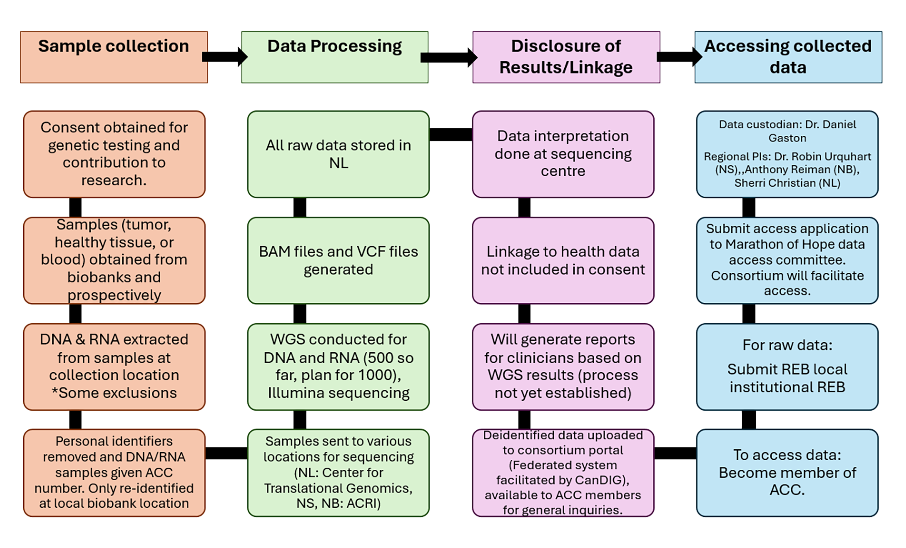
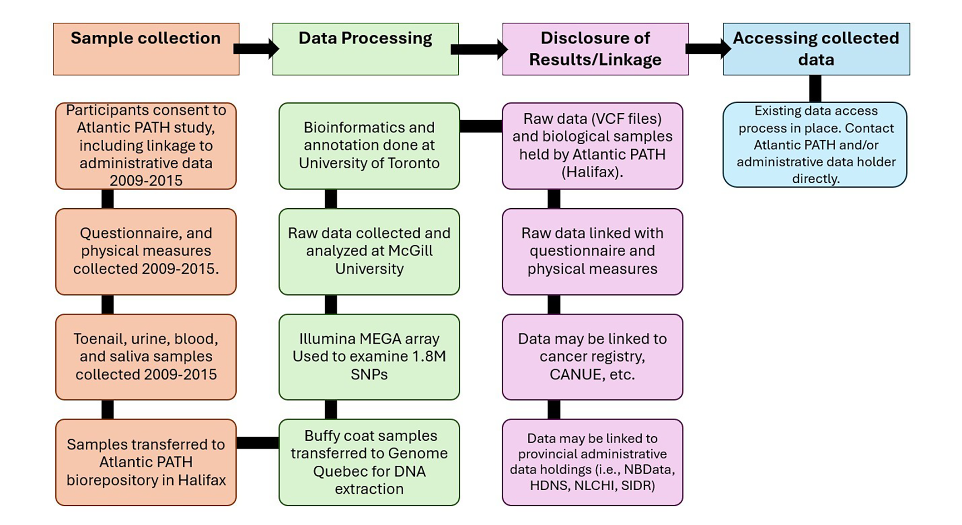
Figure 3: New Brunswick, Vitalité Health Network — Flow of Genomic Data Collected in Clinical Setting
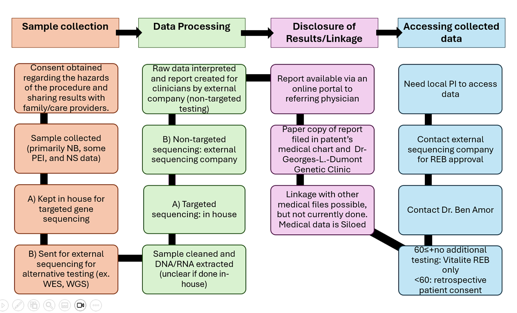
Figure 4: New Brunswick, Vitalité Health Network — Flow of Genomic Data Collected for Carrier Screening Project
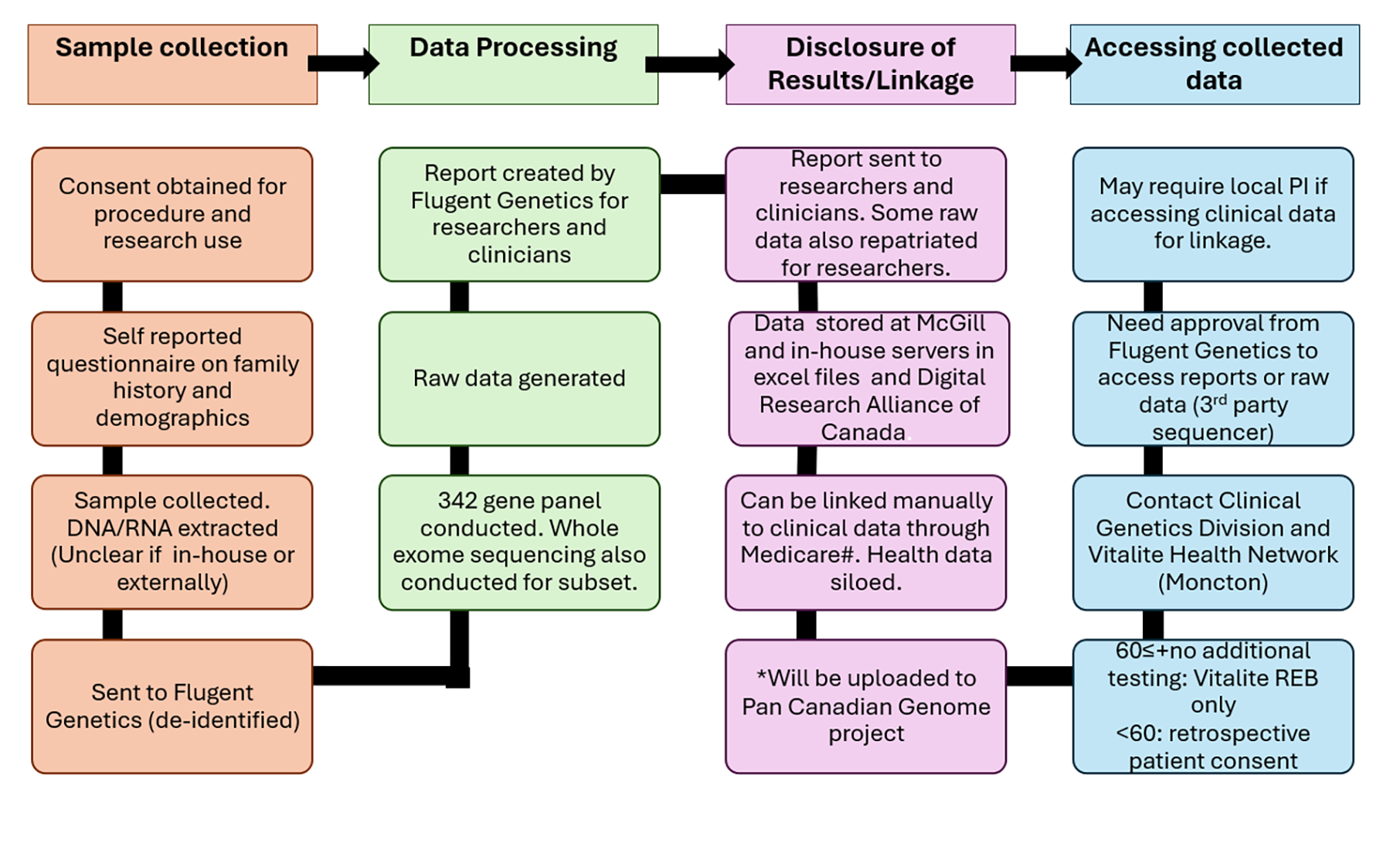
Figure 5: New Brunswick, Horizon Health Network — Flow of Genomic Data Collected in Clinical Setting
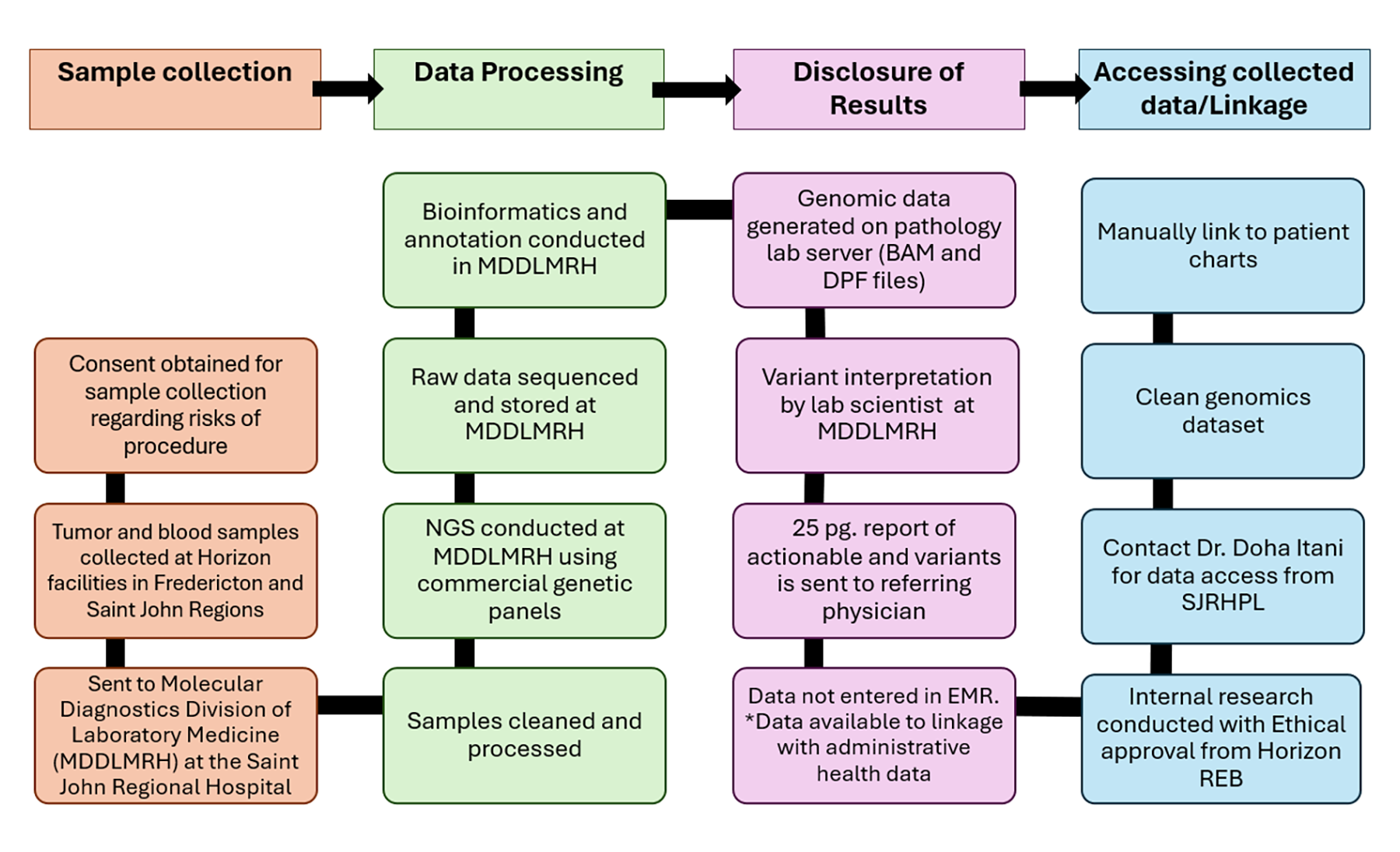
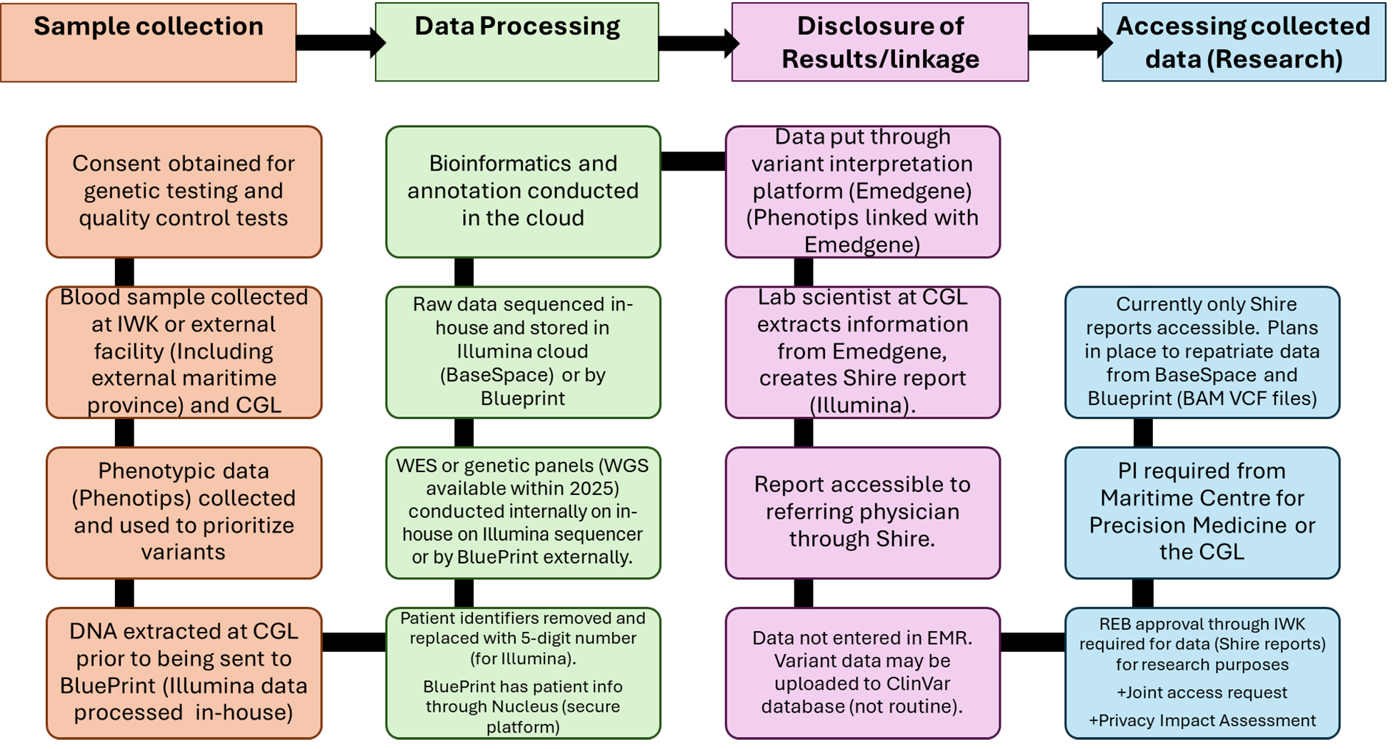
Figure 7: Nova Scotia Health Authority — Flow of Genomic Data Collected in Clinical Setting With Somatic Data
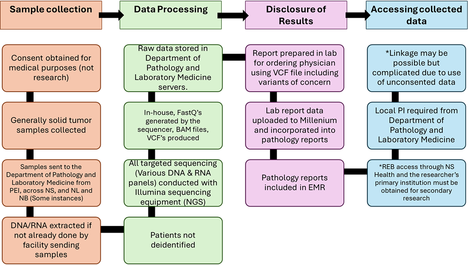
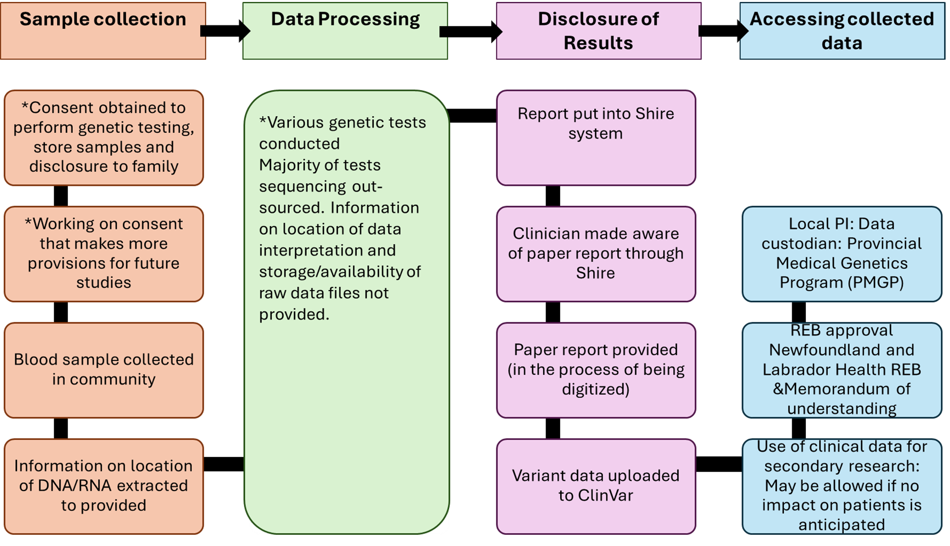
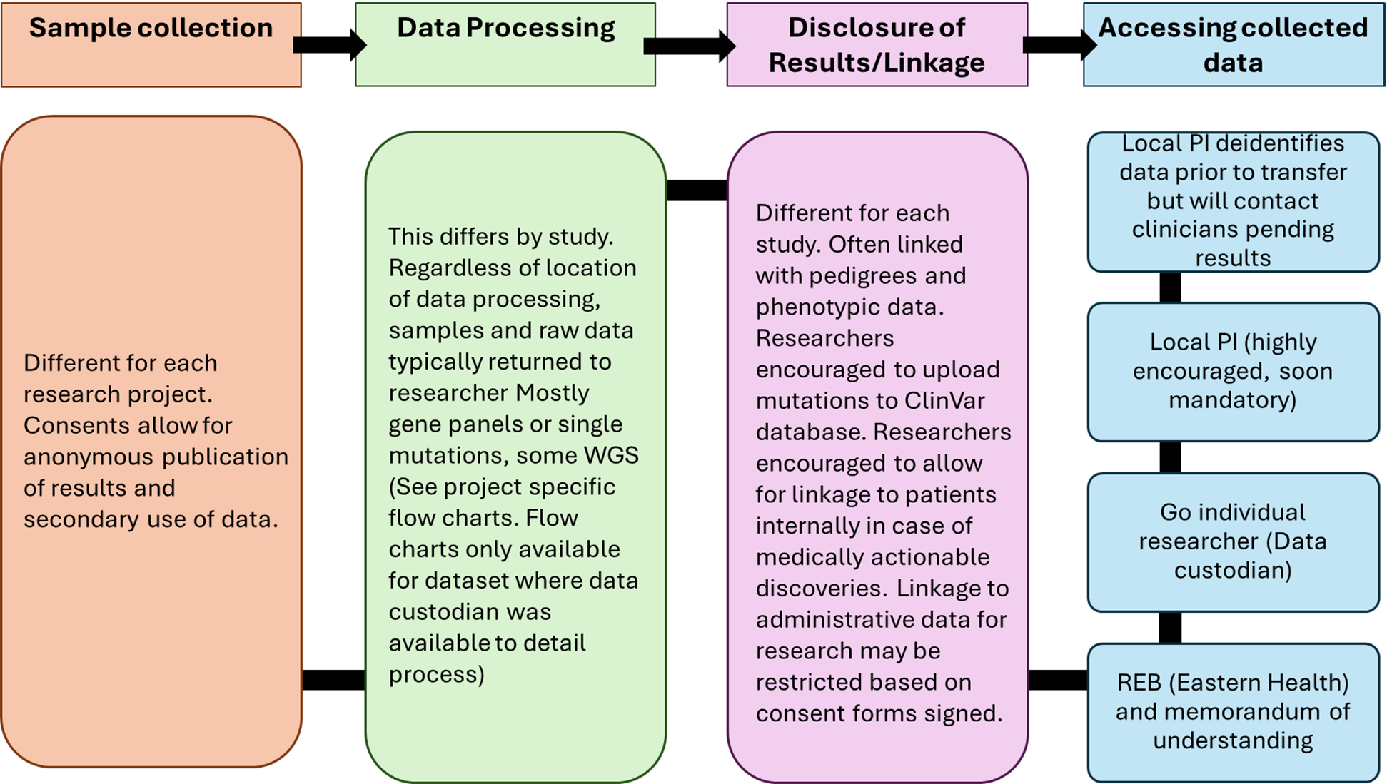
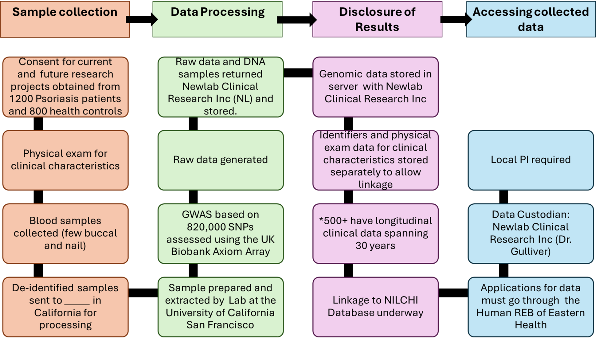
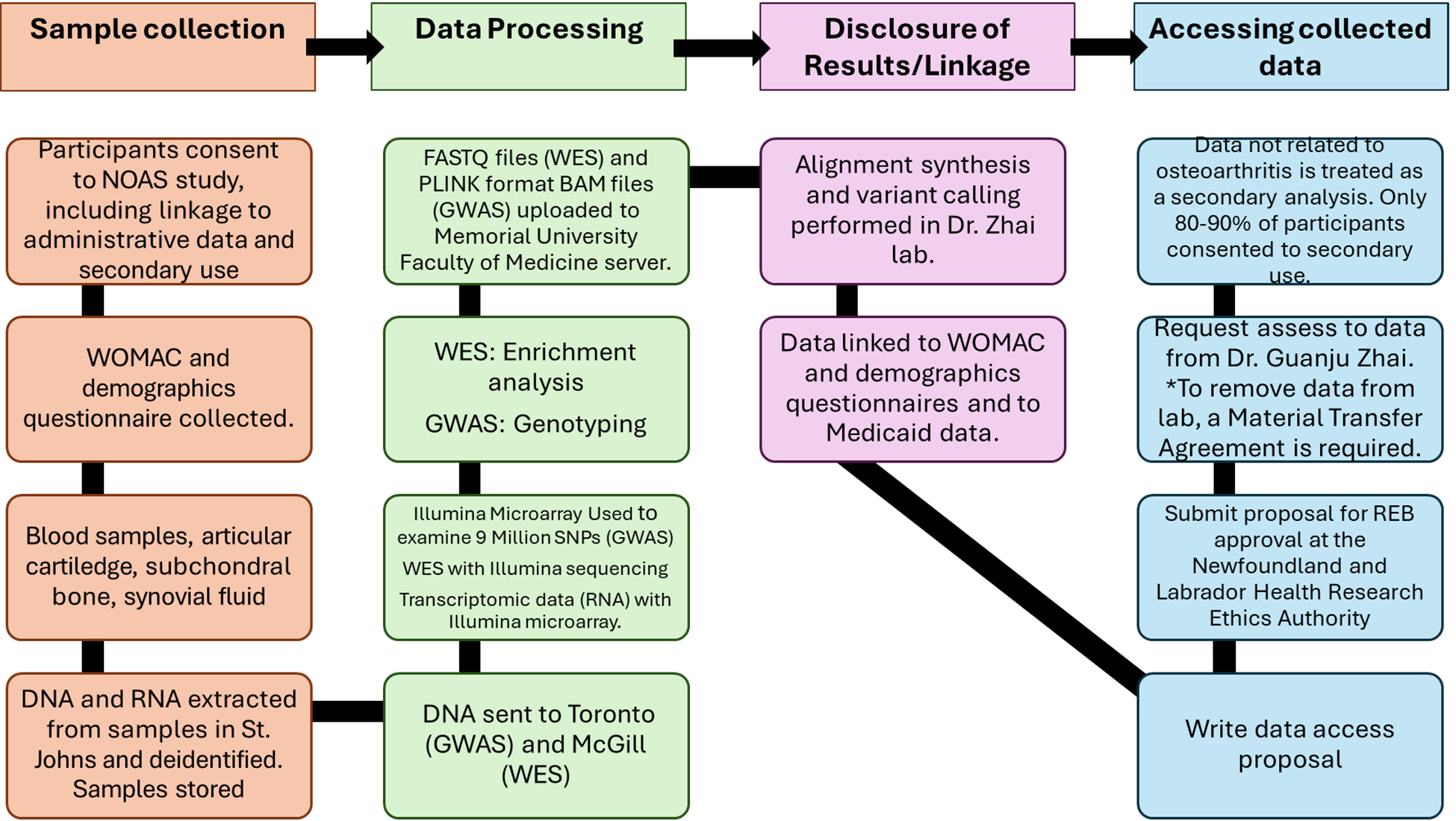
References
1.McKee-Muir O, Dyack S, Taillon M, Brock JA, Sheriko J. Epidemiology of spinal muscular atrophy caused by SMN1 deletions in Maritime Canada. Am J Med Genet A. 2023 Nov;191(11):2711–5. PubMed
2.Sweeney E, Cui Y, DeClercq V, Devichand P, Forbes C, Grandy S, et al. Cohort Profile: The Atlantic Partnership for Tomorrow’s Health (Atlantic PATH) Study. Int J Epidemiol. 2017 Dec 1;46(6):1762–1763i. PubMed
3.Levesque M, Wood R, Carter MD, Brock JA, Kieser K. Screening and testing practices for Lynch syndrome in Nova Scotians with endometrial cancer: a descriptive study. CMAJ Open. 2023 Oct;11(5):E1012–9. PubMed
4.Allen VM, Schollenberg E, Aberg E, Brock JAK. Use of Clinically Informed Strategies and Diagnostic Yields of Genetic Testing for Fetal Structural Anomalies Following a Non-Diagnostic Microarray Result: A Population-Based Cohort Study. Prenat Diagn. 2025 Mar;45(3):318–25. PubMed
5.Guernsey DL, Matsuoka M, Jiang H, Evans S, Macgillivray C, Nightingale M, et al. Mutations in origin recognition complex gene ORC4 cause Meier-Gorlin syndrome. Nat Genet. 2011 Feb 27;43(4):360–4. PubMed
6.Kokorovic A, Thomas A, Serrano-Lomelin J, Ferguson M, Rendon RA. Clinical predictors of a positive test result in patients undergoing genetic evaluation for a hereditary kidney cancer syndrome. Can Urol Assoc J. 2020 Aug;14(8):274–80. PubMed
7.Walsh NM. Primary neuroendocrine (Merkel cell) carcinoma of the skin: morphologic diversity and implications thereof. Hum Pathol. 2001 July;32(7):680–9. PubMed
8.Ly TY, Walsh NM, Pasternak S. The spectrum of Merkel cell polyomavirus expression in Merkel cell carcinoma, in a variety of cutaneous neoplasms, and in neuroendocrine carcinomas from different anatomical sites. Hum Pathol. 2012 Apr;43(4):557–66. PubMed
9.Fleming KE, Ly TY, Pasternak S, Godlewski M, Doucette S, Walsh NM. Support for p63 expression as an adverse prognostic marker in Merkel cell carcinoma: report on a Canadian cohort. Hum Pathol. 2014 May;45(5):952–60. PubMed
10.Carter MD, Gaston D, Huang WY, Greer WL, Pasternak S, Ly TY, et al. Genetic profiles of different subsets of Merkel cell carcinoma show links between combined and pure MCPyV-negative tumors. Hum Pathol. 2018 Jan;71:117–25. PubMed
11.DeCoste RC, Carter MD, Pasternak S, Fleming KE, Gaston D, Legge A, et al. Relationship between p63 and p53 expression in Merkel cell carcinoma and corresponding abnormalities in TP63 and TP53: a study and a proposal. Hum Pathol. 2021 Nov;117:31–41. PubMed
12.DeCoste RC, Walsh NM, Gaston D, Ly TY, Pasternak S, Cutler S, et al. RB1-deficient squamous cell carcinoma: the proposed source of combined Merkel cell carcinoma. Mod Pathol Off J U S Can Acad Pathol Inc. 2022 Dec;35(12):1829–36. PubMed
13.Li Z, Wang Z, Lee MC, Zenkel M, Peh E, Ozaki M, et al. Association of Rare CYP39A1 Variants With Exfoliation Syndrome Involving the Anterior Chamber of the Eye. JAMA. 2021 Feb 23;325(8):753–64. PubMed
14.Aung T, Ozaki M, Lee MC, Schlötzer-Schrehardt U, Thorleifsson G, Mizoguchi T, et al. Genetic association study of exfoliation syndrome identifies a protective rare variant at LOXL1 and five new susceptibility loci. Nat Genet. 2017 July;49(7):993–1004. PubMed
15.Cutler SD, Knopf P, Campbell CJV, Thoni A, Abou El Hassan M, Forward N, et al. DMG26: A Targeted Sequencing Panel for Mutation Profiling to Address Gaps in the Prognostication of Multiple Myeloma. J Mol Diagn JMD. 2021 Dec;23(12):1699–714. PubMed
16.Robitaille J, MacDonald MLE, Kaykas A, Sheldahl LC, Zeisler J, Dubé MP, et al. Mutant frizzled-4 disrupts retinal angiogenesis in familial exudative vitreoretinopathy. Nat Genet. 2002 Oct;32(2):326–30. PubMed
17.Ells A, Guernsey DL, Wallace K, Zheng B, Vincer M, Allen A, et al. Severe retinopathy of prematurity associated with FZD4 mutations. Ophthalmic Genet. 2010 Mar;31(1):37–43. PubMed
18.Robitaille JM, Gillett RM, LeBlanc MA, Gaston D, Nightingale M, Mackley MP, et al. Phenotypic overlap between familial exudative vitreoretinopathy and microcephaly, lymphedema, and chorioretinal dysplasia caused by KIF11 mutations. JAMA Ophthalmol. 2014 Dec;132(12):1393–9. PubMed
19.van der Ende S, Bedard K, Wallace K, Mackley MP, Nightingale M, Gaston D, et al. Gene Variant Spectrum in Probands With Familial Exudative Vitreoretinopathy Using an Expanded Panel. Invest Ophthalmol Vis Sci. 2025 Feb 3;66(2):23. PubMed
20.Sinha N, Gaston D, Manders D, Goudie M, Matsuoka M, Xie T, et al. Characterization of genome-wide copy number aberrations in colonic mixed adenoneuroendocrine carcinoma and neuroendocrine carcinoma reveals recurrent amplification of PTGER4 and MYC genes. Hum Pathol. 2018 Mar;73:16–25. PubMed
21.Hou L, Heilbronner U, Degenhardt F, Adli M, Akiyama K, Akula N, et al. Genetic variants associated with response to lithium treatment in bipolar disorder: a genome-wide association study. Lancet Lond Engl. 2016 Mar 12;387(10023):1085–93. PubMed
22.Stone W, Nunes A, Akiyama K, Akula N, Ardau R, Aubry JM, et al. Prediction of lithium response using genomic data. Sci Rep. 2021 Jan 13;11(1):1155. PubMed
23.O’Connell KS, Koromina M, van der Veen T, Boltz T, David FS, Yang JMK, et al. Genomics yields biological and phenotypic insights into bipolar disorder. Nature. 2025 Mar;639(8056):968–75. PubMed
24.McAusland L, Burton CL, Bagnell A, Boylan K, Hatchard T, Lingley-Pottie P, et al. The genetic architecture of youth anxiety: a study protocol. BMC Psychiatry. 2024 Feb 23;24(1):159. PubMed
25.Kmochová T, Kidd KO, Orr A, Hnízda A, Hartmannová H, Hodaňová K, et al. Autosomal dominant ApoA4 mutations present as tubulointerstitial kidney disease with medullary amyloidosis. Kidney Int. 2024 Apr;105(4):799–811. PubMed
26.Ouellette RJ, Richard D, Maïcas E. RT-PCR for mammaglobin genes, MGB1 and MGB2, identifies breast cancer micrometastases in sentinel lymph nodes. Am J Clin Pathol. 2004 May;121(5):637–43. PubMed
27.Gauvin K, Allain V, Bouhamdani N, Williams C, Saheb Y, Savoie C, et al. Retrospective Study of Genetic Testing Results Reveals Pathogenic Variants Beyond BRCA1/2 in Hereditary Breast and Ovarian Cancer Cases in New Brunswick: Implications for Future Care. Cancer Med. 2025 Feb;14(3):e70640. PubMed
28.Northrup V, Maybank A, Carson N, Rahmeh T. The Value of Next-Generation Sequencing in the Screening and Evaluation of Hematologic Neoplasms in Clinical Practice. Am J Clin Pathol. 2020 Apr 15;153(5):639–45. PubMed
29.Robichaud PP, Allain EP, Belbraouet S, Bhérer C, Mamelona J, Harquail J, et al. Pathogenic variants carrier screening in New Brunswick: Acadians reveal high carrier frequency for multiple genetic disorders. BMC Med Genomics. 2022 Dec;15(1):98. PubMed
30.Roy JW, Wajnberg G, Ouellette A, Boucher JE, Lacroix J, Chacko S, et al. Small RNA sequencing analysis of peptide-affinity isolated plasma extracellular vesicles distinguishes pancreatic cancer patients from non-affected individuals. Sci Rep. 2023 June 7;13(1):9251. PubMed
31.Zurel H, Bhérer C, Batten R, MacMillan ME, Demiriz S, Mirhendi S, et al. Characterization of Y chromosome diversity in newfoundland and labrador: evidence for a structured founding population. Eur J Hum Genet. 2025 Jan;33(1):98–107. PubMed
32.Gilbert E, Zurel H, MacMillan ME, Demiriz S, Mirhendi S, Merrigan M, et al. The Newfoundland and Labrador mosaic founder population descends from an Irish and British diaspora from 300 years ago. Commun Biol. 2023 Apr 28;6(1):469. PubMed
33.Rahman P, Jones A, Curtis J, Bartlett S, Peddle L, Fernandez BA, et al. The Newfoundland population: a unique resource for genetic investigation of complex diseases. Hum Mol Genet. 2003 Oct 15;12(suppl 2):R167–72. PubMed
34.Zhai G, Zhou J, Woods MO, Green JS, Parfrey P, Rahman P, et al. Genetic structure of the Newfoundland and Labrador population: founder effects modulate variability. Eur J Hum Genet. 2016 July;24(7):1063–70. PubMed
35.Zipperlen K, Peddle L, Melay B, Hefferton D, Rahman P. Association of TNF-α polymorphisms in Crohn disease. Hum Immunol. 2005 Jan;66(1):56–9. PubMed
36.Gladman DD, Farewell VT, Pellett F, Schentag C, Rahman P. HLA is a candidate region for psoriatic arthritis. Hum Immunol. 2003 Sept;64(9):887–9. PubMed
37.Butt C, Sun S, Greenwood C, Gladman D, Rahman P. Lack of association of SLC22A4, SLC22A5, SLC9A3R1 and RUNX1 variants in psoriatic arthritis. Rheumatology. 2005 June 1;44(6):820–1. PubMed
38.Butt C, Sun S, Peddle L, Greenwood C, Hamilton S, Gladman D, et al. Association of nuclear factor-kappaB in psoriatic arthritis. J Rheumatol. 2005 Sept;32(9):1742–4. PubMed
39.Butt C, Peddle L, Greenwood C, Hamilton S, Gladman D, Rahman P. Association of functional variants of PTPN22 and tp53in psoriatic arthritis: a case-control study. Arthritis Res Ther. 2006 Jan 3;8(1):R27. PubMed
40.Rahman P, Siannis F, Butt C, Farewell V, Peddle L, Pellett F, et al. TNFα polymorphisms and risk of psoriatic arthritis. Ann Rheum Dis. 2006 July;65(7):919–23. PubMed
41.Rahman P, Sun S, Peddle L, Snelgrove T, Melay W, Greenwood C, et al. Association between the interleukin‐1 family gene cluster and psoriatic arthritis. Arthritis Rheum. 2006 July;54(7):2321–5. PubMed
42.Butt C, Lim S, Greenwood C, Rahman P. VEGF, FGF1, FGF2 and EGF gene polymorphisms and psoriatic arthritis. BMC Musculoskelet Disord. 2007 Dec;8(1):1. PubMed
43.Rahman P, Inman RD, Maksymowych WP, Reeve JP, Peddle L, Gladman DD. Association of Interleukin 23 Receptor Variants with Psoriatic Arthritis. J Rheumatol. 2009 Jan;36(1):137–40. PubMed
44.Pollock RA, Chandran V, Pellett FJ, Thavaneswaran A, Eder L, Barrett J, et al. The functional MICA ‐129 polymorphism is associated with skin but not joint manifestations of psoriatic disease independently of HLA ‐B and HLA ‐C. Tissue Antigens. 2013 July;82(1):43–7. PubMed
45.O’Rielly DD, Pollock R, Zhang Y, Al-Ghanim N, Yazdani R, Hamilton S, et al. AB0160 Epigenetic Studies in Maternally versus Paternally Transmitted Psoriatic Disease. Ann Rheum Dis. 2014 June;73:856.
46.Chandran V, O’Reilly, Daniel, Haddad, Ayat, Zhang, Yufeng, Zhai, Guangju, Codner, Douglas. Genome-Wide Methylome Investigation Reveals New Candidate Genes Associated with Arthritis Mutilans. Arthritis Rheumatol. 2014;66(Supplement 10):S274.
47.O’Rielly DD, Zhang Y, Codner D, Dohey A, Zhou A, Al Ghanim N, et al. OP0200 Global DNA Methylation Patterns Differ Between Responders and Non-Responders in Psoriatic Arthritis Patients Treated with Tumor Necrosis Factor-α Inhibitors. Ann Rheum Dis. 2015 June;74:147.
48.Pollock RA, Thavaneswaran A, Pellett F, Chandran V, Petronis A, Rahman P, et al. Further Evidence Supporting a Parent‐of‐Origin Effect in Psoriatic Disease. Arthritis Care Res. 2015 Nov;67(11):1586–90. PubMed
49.Stuart PE, Nair RP, Ellinghaus E, Ding J, Tejasvi T, Gudjonsson JE, et al. Genome-wide association analysis identifies three psoriasis susceptibility loci. Nat Genet. 2010 Nov;42(11):1000–4. PubMed
50.Stuart PE, Nair RP, Tsoi LC, Tejasvi T, Das S, Kang HM, et al. Genome-wide Association Analysis of Psoriatic Arthritis and Cutaneous Psoriasis Reveals Differences in Their Genetic Architecture. Am J Hum Genet. 2015 Dec;97(6):816–36. PubMed
51.Rahmati S, O’Rielly DD, Li Q, Codner D, Dohey A, Jenkins K, et al. Rho-GTPase pathways may differentiate treatment response to TNF-alpha and IL-17A inhibitors in psoriatic arthritis. Sci Rep. 2020 Dec 10;10(1):21703. PubMed
52.Nair RP, Duffin KC, Helms C, Ding J, Stuart PE, Goldgar D, et al. Genome-wide scan reveals association of psoriasis with IL-23 and NF-κB pathways. Nat Genet. 2009 Feb;41(2):199–204. PubMed
53.Das S, Stuart PE, Ding J, Tejasvi T, Li Y, Tsoi LC, et al. Fine mapping of eight psoriasis susceptibility loci. Eur J Hum Genet. 2015 June;23(6):844–53. PubMed
54.O’Rielly DD, Uddin M, Codner D, Hayley M, Zhou J, Pena-Castillo L, et al. Private rare deletions in SEC16A and MAMDC4 may represent novel pathogenic variants in familial axial spondyloarthritis. Ann Rheum Dis. 2016 Apr;75(4):772–9. PubMed
55.Lehr J, Rahman P, O’Rielly DD. High Accuracy and Significant Savings Using Tag-SNP Genotyping to Determine HLA-B*27 Status. J Rheumatol. 2017 June;44(6):962.2-963.
56.Spector TD, Reneland RH, Mah S, Valdes AM, Hart DJ, Kammerer S, et al. Association between a variation in LRCH1 and knee osteoarthritis: A genome‐wide single‐nucleotide polymorphism association study using DNA pooling. Arthritis Rheum. 2006 Feb;54(2):524–32. PubMed
57.Aref-Eshghi E, Zhang Y, Liu M, Harper PE, Martin G, Furey A, et al. Genome-wide DNA methylation study of hip and knee cartilage reveals embryonic organ and skeletal system morphogenesis as major pathways involved in osteoarthritis. BMC Musculoskelet Disord. 2015 Dec;16(1):287. PubMed
58.Gill R, Liu M, Sun G, Furey A, Spector T, Rahman P, et al. Genomic heterozygosity is associated with a lower risk of osteoarthritis. BMC Genomics. 2024 Jan 20;25(1):85. PubMed
59.Zhai G, Aref-Eshghi E, Zhang H, Martin G, Furey A, Sun G. Attempt to replicate the published osteoarthritis-associated genetic variants in the Newfoundland & Labrador Population. J Orthop Rheumatol. 2014;1(3).
60.Aref-Eshghi E, Liu M, Harper PE, Doré J, Martin G, Furey A, et al. Overexpression of MMP13 in human osteoarthritic cartilage is associated with the SMAD-independent TGF-β signalling pathway. Arthritis Res Ther. 2015 Dec;17(1):264. PubMed
61.Aref-Eshghi E, Liu M, Razavi-Lopez SB, Hirasawa K, Harper PE, Martin G, et al. SMAD3 Is Upregulated in Human Osteoarthritic Cartilage Independent of the Promoter DNA Methylation. J Rheumatol. 2016 Feb;43(2):388–94. PubMed
62.Werdyani, S., Liu, M., Xie, Z., Furey, A., Gao, Z., Rahman, P., et al. Genes related to muscle strength, behavioural trait, pain response, and inflammation are associated with poor outcome of the total joint replacement therapy in primary osteoarthritis patients. 2021;103.
63.Werdyani, S., Liu, M., Furey, A., Gao, Z., Rahman, P., Zhai, G. A genome-wide association study identified novel genes associated with osteoarthritis. 2022;30:S132.
64.Maksymowych WP, Rahman P, Reeve JP, Gladman DD, Peddle L, Inman RD. Association of the IL1 gene cluster with susceptibility to ankylosing spondylitis: An analysis of three Canadian populations. Arthritis Rheum. 2006 Mar;54(3):974–85. PubMed
65.Snelgrove T, Lim S, Greenwood C, Peddle L, Hamilton S, Inman R, et al. Association of toll-like receptor 4 variants and ankylosing spondylitis: a case-control study. J Rheumatol. 2007 Feb;34(2):368–70. PubMed
66.Rahman P, Inman RD, Gladman DD, Reeve JP, Peddle L, Maksymowych WP. Association of interleukin‐23 receptor variants with ankylosing spondylitis. Arthritis Rheum. 2008 Apr;58(4):1020–5. PubMed
67.Maksymowych WP, Inman RD, Gladman DD, Reeve JP, Pope A, Rahman P. Association of a specific ERAP1/ARTS1 haplotype with disease susceptibility in ankylosing spondylitis. Arthritis Rheum. 2009 May;60(5):1317–23. PubMed
68.Uddin M, Maksymowych WP, Inman R, Gladman D, Munn A, Yazdani R, et al. UGT2B17 copy number gain in a large ankylosing spondylitis multiplex family. BMC Genet. 2013 Dec;14(1):67. PubMed
69.Young TL. Non-syndromic progressive hearing loss DFNA38 is caused by heterozygous missense mutation in the Wolfram syndrome gene WFS1. Hum Mol Genet. 2001 Oct 1;10(22):2509–14. PubMed
70.Ahmed ZM, Cindy Li X, Powell SD, Riazuddin S, Young TL, Ramzan K, et al. Characterization of a new full length TMPRSS3 isoform and identification of mutant alleles responsible for nonsyndromic recessive deafness in Newfoundland and Pakistan. BMC Med Genet. 2004 Dec;5(1):24. PubMed
71.Abdelfatah N, McComiskey DA, Doucette L, Griffin A, Moore SJ, Negrijn C, et al. Identification of a novel in-frame deletion in KCNQ4 (DFNA2A) and evidence of multiple phenocopies of unknown origin in a family with ADSNHL. Eur J Hum Genet. 2013 Oct;21(10):1112–9. PubMed
72.Pater JA, Benteau T, Griffin A, Penney C, Stanton SG, Predham S, et al. A common variant in CLDN14 causes precipitous, prelingual sensorineural hearing loss in multiple families due to founder effect. Hum Genet. 2017 Jan;136(1):107–18. PubMed
73.Pater JA, Penney C, O’Rielly DD, Griffin A, Kamal L, Brownstein Z, et al. Autosomal dominant non-syndromic hearing loss maps to DFNA33 (13q34) and co-segregates with splice and frameshift variants in ATP11A, a phospholipid flippase gene. Hum Genet. 2022 Apr;141(3–4):431–44. PubMed
74.Abdelfatah N, Mostafa AA, French CR, Doucette LP, Penney C, Lucas MB, et al. A pathogenic deletion in Forkhead Box L1 (FOXL1) identifies the first otosclerosis (OTSC) gene. Hum Genet. 2022 Apr;141(3–4):965–79. PubMed
75.Singh S, Penney C, Griffin A, Woodland G, Werdyani S, Benteau TA, et al. Highly variable hearing loss due to POU4F3 (c.37del) is revealed by longitudinal, frequency specific analyses. Eur J Hum Genet. 2023 July;31(7):815–23. PubMed
76.Green JS, O’Rielly DD, Pater JA, Houston J, Rajabi H, Galutira D, et al. The genetic architecture of Stargardt macular dystrophy (STGD1): a longitudinal 40-year study in a genetic isolate. Eur J Hum Genet. 2020 July;28(7):925–37. PubMed
77.Fan Y, Rahman P, Peddle L, Hefferton D, Gladney N, Moore SJ, et al. Bardet–Biedl syndrome 1 genotype and obesity in the Newfoundland population. Int J Obes. 2004 May;28(5):680–4. PubMed
78.Service S, DeYoung J, Karayiorgou M, Roos JL, Pretorious H, Bedoya G, et al. Magnitude and distribution of linkage disequilibrium in population isolates and implications for genome-wide association studies. Nat Genet. 2006 May;38(5):556–60. PubMed
79.Aksentijevich I, Masters SL, Ferguson PJ, Dancey P, Frenkel J, Van Royen-Kerkhoff A, et al. An Autoinflammatory Disease with Deficiency of the Interleukin-1–Receptor Antagonist. N Engl J Med. 2009 June 4;360(23):2426–37. PubMed
80.Dawson LM, Smith KN, Werdyani S, Ndikumana R, Penney C, Wiede LL, et al. A dominant RAD51C pathogenic splicing variant predisposes to breast and ovarian cancer in the Newfoundland population due to founder effect. Mol Genet Genomic Med. 2020 Feb;8(2):e1070. PubMed
81.Pedram P, Zhai G, Gulliver W, Zhang H, Sun G. Two novel candidate genes identified in adults from the Newfoundland population with addictive tendencies towards food. Appetite. 2017 Aug;115:71–9. PubMed
82.Young TL, Woods MO, Parfrey PS, Green JS, O’Leary E, Hefferton D, et al. Canadian Bardet-Biedl syndrome family reduces the critical region of BBS3 (3p) and presents with a variable phenotype. Am J Med Genet. 1998 Aug 6;78(5):461–7. PubMed
83.Woods MO, Young TL, Parfrey PS, Hefferton D, Green JS, Davidson WS. Genetic Heterogeneity of Bardet–Biedl Syndrome in a Distinct Canadian Population: Evidence for a Fifth Locus. Genomics. 1999 Jan;55(1):2–9. PubMed
84.Young TL, Woods MO, Parfrey PS, Green JS, Hefferton D, Davidson WS. A Founder Effect in the Newfoundland Population Reduces the Bardet-Biedl Syndrome I (BBS1) Interval to 1 cM. Am J Hum Genet. 1999 Dec;65(6):1680–7. PubMed
85.Katsanis N, Beales PL, Woods MO, Lewis RA, Green JS, Parfrey PS, et al. Mutations in MKKS cause obesity, retinal dystrophy and renal malformations associated with Bardet-Biedl syndrome. Nat Genet. 2000 Sept;26(1):67–70. PubMed
86.Beales PL, Katsanis N, Lewis RA, Ansley SJ, Elcioglu N, Raza J, et al. Genetic and Mutational Analyses of a Large Multiethnic Bardet-Biedl Cohort Reveal a Minor Involvement of BBS6 and Delineate the Critical Intervals of Other Loci. Am J Hum Genet. 2001 Mar;68(3):606–16. PubMed
87.Moore SJ, Green JS, Fan Y, Bhogal AK, Dicks E, Fernandez BA, et al. Clinical and genetic epidemiology of Bardet-Biedl syndrome in Newfoundland: A 22-year prospective, population-based, cohort study. Am J Med Genet A. 2005 Feb 1;132A(4):352–60. PubMed
88.Stuckless S, Parfrey PS, Woods MO, Cox J, Fitzgerald GW, Green JS, et al. The phenotypic expression of three MSH2 mutations in large Newfoundland families with Lynch syndrome. Fam Cancer. 2007 Feb 13;6(1):1–12. PubMed
89.Campbell PT, Edwards L, McLaughlin JR, Green J, Younghusband HB, Woods MO. Cytochrome P450 17A1 and Catechol O -Methyltransferase Polymorphisms and Age at Lynch Syndrome Colon Cancer Onset in Newfoundland. Clin Cancer Res. 2007 July 1;13(13):3783–8. PubMed
90.Woods MO, Williams P, Careen A, Edwards L, Bartlett S, McLaughlin JR, et al. A new variant database for mismatch repair genes associated with Lynch syndrome. Hum Mutat. 2007 July;28(7):669–73. PubMed
91.Kohonen‐Corish MRJ, Macrae F, Genuardi M, Aretz S, Bapat B, Bernstein IT, et al. Deciphering the colon cancer genes—report of the InSiGHT‐Human Variome Project Workshop, UNESCO, Paris 2010. Hum Mutat. 2011 Apr;32(4):491–4. PubMed
92.Plazzer JP, Sijmons RH, Woods MO, Peltomäki P, Thompson B, Den Dunnen JT, et al. The InSiGHT database: utilizing 100 years of insights into Lynch Syndrome. Fam Cancer. 2013 June;12(2):175–80. PubMed
93.Thompson BA, Spurdle AB, Plazzer JP, Greenblatt MS, Akagi K, Al-Mulla F, et al. Application of a 5-tiered scheme for standardized classification of 2,360 unique mismatch repair gene variants in the InSiGHT locus-specific database. Nat Genet. 2014 Feb;46(2):107–15. PubMed
94.Win AK, Dowty JG, Reece JC, Lee G, Templeton AS, Plazzer JP, et al. Variation in the risk of colorectal cancer in families with Lynch syndrome: a retrospective cohort study. Lancet Oncol. 2021 July;22(7):1014–22. PubMed
95.Woods MO, Hyde AJ, Curtis FK, Stuckless S, Green JS, Pollett AF, et al. High Frequency of Hereditary Colorectal Cancer in Newfoundland Likely Involves Novel Susceptibility Genes. Clin Cancer Res. 2005 Oct 1;11(19):6853–61. PubMed
96.Wish TA, Hyde AJ, Parfrey PS, Green JS, Younghusband HB, Simms MI, et al. Increased Cancer Predisposition in Family Members of Colorectal Cancer Patients Harboring the p.V600E BRAF Mutation: a Population-Based Study. Cancer Epidemiol Biomarkers Prev. 2010 July 1;19(7):1831–9. PubMed
97.Woods MO, Younghusband HB, Parfrey PS, Gallinger S, McLaughlin J, Dicks E, et al. The genetic basis of colorectal cancer in a population-based incident cohort with a high rate of familial disease. Gut. 2010 Oct 1;59(10):1369–77. PubMed
98.Clarke E, Green RC, Green JS, Mahoney K, Parfrey PS, Younghusband HB, et al. Inherited deleterious variants in GALNT12 are associated with CRC susceptibility. Hum Mutat. 2012 July;33(7):1056–8. PubMed
99.DeRycke MS, Gunawardena SR, Middha S, Asmann YW, Schaid DJ, McDonnell SK, et al. Identification of Novel Variants in Colorectal Cancer Families by High-Throughput Exome Sequencing. Cancer Epidemiol Biomarkers Prev. 2013 July 1;22(7):1239–51. PubMed
100.Haja Mohideen AMS, Hyde A, Squires J, Wang J, Dicks E, Younghusband B, et al. Examining the Polymorphisms in the Hypoxia Pathway Genes in Relation to Outcome in Colorectal Cancer. PLoS ONE. 2014 Nov 18;9(11):e113513.
101.Haja Mohideen AMS, Dicks E, Parfrey P, Green R, Savas S. Mitochondrial DNA polymorphisms, its copy number change and outcome in colorectal cancer. BMC Res Notes. 2015 Dec;8(1):272. PubMed
102.Xu W, Xu J, Shestopaloff K, Dicks E, Green J, Parfrey P, et al. A genome wide association study on Newfoundland colorectal cancer patients’ survival outcomes. Biomark Res. 2015 Dec;3(1):6. PubMed
103.Zhu Y, Wang PP, Zhai G, Bapat B, Savas S, Woodrow JR, et al. Vitamin D receptor and calcium-sensing receptor polymorphisms and colorectal cancer survival in the Newfoundland population. Br J Cancer. 2017 Sept;117(6):898–906. PubMed
104.Zhu Y, Wang PP, Zhai G, Bapat B, Savas S, Woodrow JR, et al. Association of rs2282679 A>C polymorphism in vitamin D binding protein gene with colorectal cancer risk and survival: effect modification by dietary vitamin D intake. BMC Cancer. 2018 Dec;18(1):155. PubMed
105.Curtis A, Yu Y, Carey M, Parfrey P, Yilmaz YE, Savas S. Examining SNP-SNP interactions and risk of clinical outcomes in colorectal cancer using multifactor dimensionality reduction based methods. Front Genet. 2022 Aug 3;13:902217. PubMed
106.Savas S, Xu J, Werdyani S, Shestopaloff K, Dicks E, Green J, et al. A Survival Association Study of 102 Polymorphisms Previously Associated with Survival Outcomes in Colorectal Cancer. BioMed Res Int. 2015;2015:1–9. PubMed
107.Dorani F, Hu T, Woods MO, Zhai G. Ensemble learning for detecting gene-gene interactions in colorectal cancer. PeerJ. 2018 Oct 29;6:e5854. PubMed
108.Schumacher FR, Schmit SL, Jiao S, Edlund CK, Wang H, Zhang B, et al. Genome-wide association study of colorectal cancer identifies six new susceptibility loci. Nat Commun. 2015 July 7;6(1):7138. PubMed
109.Bien SA, Su YR, Conti DV, Harrison TA, Qu C, Guo X, et al. Genetic variant predictors of gene expression provide new insight into risk of colorectal cancer. Hum Genet. 2019 Apr;138(4):307–26. PubMed
110.Guo X, Lin W, Wen W, Huyghe J, Bien S, Cai Q, et al. Identifying Novel Susceptibility Genes for Colorectal Cancer Risk From a Transcriptome-Wide Association Study of 125,478 Subjects. Gastroenterology. 2021 Mar;160(4):1164-1178.e6. PubMed
111.Thomas M, Sakoda LC, Hoffmeister M, Rosenthal EA, Lee JK, Van Duijnhoven FJB, et al. Genome-wide Modeling of Polygenic Risk Score in Colorectal Cancer Risk. Am J Hum Genet. 2020 Sept;107(3):432–44. PubMed
112.Archambault AN, Su YR, Jeon J, Thomas M, Lin Y, Conti DV, et al. Cumulative Burden of Colorectal Cancer–Associated Genetic Variants Is More Strongly Associated With Early-Onset vs Late-Onset Cancer. Gastroenterology. 2020 Apr;158(5):1274-1286.e12. PubMed
113.Archambault AN, Jeon J, Lin Y, Thomas M, Harrison TA, Bishop DT, et al. Risk Stratification for Early-Onset Colorectal Cancer Using a Combination of Genetic and Environmental Risk Scores: An International Multi-Center Study. JNCI J Natl Cancer Inst. 2022 Jan 13;djac003. PubMed
114.Schmit SL, Edlund CK, Schumacher FR, Gong J, Harrison TA, Huyghe JR, et al. Novel Common Genetic Susceptibility Loci for Colorectal Cancer. JNCI J Natl Cancer Inst. 2019 Feb 1;111(2):146–57. PubMed
115.Huyghe JR, Bien SA, Harrison TA, Kang HM, Chen S, Schmit SL, et al. Discovery of common and rare genetic risk variants for colorectal cancer. Nat Genet. 2019 Jan;51(1):76–87. PubMed
116.Huyghe JR, Harrison TA, Bien SA, Hampel H, Figueiredo JC, Schmit SL, et al. Genetic architectures of proximal and distal colorectal cancer are partly distinct. Gut. 2021 July;70(7):1325–34. PubMed
117.Savas S, Hyde A, Stuckless SN, Parfrey P, Younghusband HB, Green R. Serotonin Transporter Gene (SLC6A4) Variations Are Associated with Poor Survival in Colorectal Cancer Patients. PLoS ONE. 2012 July 24;7(7):e38953.
118.Dan LA, Werdyani S, Xu J, Shestopaloff K, Hyde A, Dicks E, et al. No associations of a set of SNPs in the Vascular Endothelial Growth Factor (VEGF) and Matrix Metalloproteinase (MMP) genes with survival of colorectal cancer patients. Cancer Med. 2016 Sept;5(9):2221–31. PubMed
119.Fernandez BA, Fox G, Bhatia R, Sala E, Noble B, Denic N, et al. A Newfoundland cohort of familial and sporadic idiopathic pulmonary fibrosis patients: clinical and genetic features. Respir Res. 2012 Dec;13(1):64. PubMed
120.Powell AE, Fernandez BA, Maroun F, Noble B, Woods MO. Familial Intracranial Aneurysm in Newfoundland: Clinical and Genetic Analysis. Can J Neurol Sci. 2019 Sept;46(5):518–26. PubMed
121.Hodgkinson KA, Parfrey PS, Bassett AS, Kupprion C, Drenckhahn J, Norman MW, et al. The impact of implantable cardioverter-defibrillator therapy on survival in autosomal-dominant arrhythmogenic right ventricular cardiomyopathy (ARVD5). J Am Coll Cardiol. 2005 Feb;45(3):400–8. PubMed
122.Merner ND, Hodgkinson KA, Haywood AFM, Connors S, French VM, Drenckhahn JD, et al. Arrhythmogenic Right Ventricular Cardiomyopathy Type 5 Is a Fully Penetrant, Lethal Arrhythmic Disorder Caused by a Missense Mutation in the TMEM43 Gene. Am J Hum Genet. 2008 Apr;82(4):809–21. PubMed
123.Haywood AFM, Merner ND, Hodgkinson KA, Houston J, Syrris P, Booth V, et al. Recurrent missense mutations in TMEM43 (ARVD5) due to founder effects cause arrhythmogenic cardiomyopathies in the UK and Canada. Eur Heart J. 2013 Apr 1;34(13):1002–11. PubMed
124.Hodgkinson KA, Connors S, Merner N, Haywood A, Young TL, McKenna W, et al. The natural history of a genetic subtype of arrhythmogenic right ventricular cardiomyopathy caused by a p.S358L mutation in TMEM43. Clin Genet. 2013 Apr;83(4):321–31. PubMed
125.Hodgkinson KA, Howes AJ, Boland P, Shen XS, Stuckless S, Young TL, et al. Long-Term Clinical Outcome of Arrhythmogenic Right Ventricular Cardiomyopathy in Individuals With a p.S358L Mutation in TMEM43 Following Implantable Cardioverter Defibrillator Therapy. Circ Arrhythm Electrophysiol. 2016 Mar;9(3):e003589. PubMed
126.Bourassa CV, Meijer IA, Merner ND, Grewal KK, Stefanelli MG, Hodgkinson K, et al. VAMP1 Mutation Causes Dominant Hereditary Spastic Ataxia in Newfoundland Families. Am J Hum Genet. 2012 Sept;91(3):548–52. PubMed
127.Abdel-Razek, O, Collier A, Predham S, Curtis S, Bullen A, Benteau T, et al. Sex-influenced mortality in three well-ascertained families with catecholaminergic polymorphic ventricular tachycardia caused by a RYR2 p.R420W mutation: The power of extended family history. Can J Cardiol. 2017;33:S96–7.
128.Etchegary H, Pike A, Puddester R, Watkins K, Warren M, Francis V, et al. Cancer prevention in cancer predisposition syndromes: A protocol for testing the feasibility of building a hereditary cancer research registry and nurse navigator follow up model. PLOS ONE. 2022 Dec 22;17(12):e0279317.
129.Dyer M, Lin QXX, Shapoval S, Thieffry D, Benoukraf T. MethMotif.Org 2024: a database integrating context-specific transcription factor-binding motifs with DNA methylation patterns. Nucleic Acids Res. 2024 Jan 5;52(D1):D222–8. PubMed
ISSN: 2563-6596
This article — unlike others published in the journal — was submitted by an author group external to Canada’s Drug Agency. It was peer reviewed by 2 external reviewers, revised by the corresponding author, and accepted for publication in the journal. Care has been taken to ensure that the information presented is original and has not been submitted for publication elsewhere.
The copyright and other intellectual property rights in this document are owned by the Canadian Agency for Drugs and Technologies in Health (operating as Canada’s Drug Agency) and its licensors and may only be used for noncommercial use, personal use, or private research and study. This material is made available for informational purposes only and no representations or warranties are made with respect to its fitness for any particular purpose. This article should not be used as a substitute for professional medical advice or the application of professional judgment in any decision-making process. Individuals may use this document at their own risk.