Drugs, Health Technologies, Health Systems
Health Technology Review
Point-of-Care Ultrasound for Guided Central Venous Catheter Insertion Compared to the Landmark Method
Key Messages
What Is the Issue?
Conventional central line insertion technique using the landmark method to identify the target vein for catheter insertion does not account for potential anatomical variations at the insertion site.
The landmark method is associated with a high risk of mechanical complications. An X-ray exam is often required to confirm correct placement to minimize complications.
Point-of-care ultrasound (POCUS) for central venous catheter (CVC) guidance has become a routine method in emergency medicine for identifying the vein location and providing real-time guidance for insertion.
A chest X-ray represents the gold standard for verifying the position of the catheter tip after it has been inserted. Although using POCUS to guide CVC insertion removes the need for an X-ray to confirm its correct placement, an X-ray is still commonly ordered after CVC placement in the intensive care unit (ICU) and emergency department settings.
The use of POCUS-guided CVC insertion continues to expand in clinical practice, yet its clinical effectiveness compared with the landmark method remains unclear.
What Did We Do?
To support decision-making, we conducted a rapid review to identify and summarize current studies and evidence-based guidelines that compared the clinical effectiveness of POCUS to the landmark method for CVC insertion. This report is an update to a previous report published in 2023.1
What Did We Find?
Current evidence suggests that the use of POCUS to guide CVC placement is effective compared with the landmark method.
POCUS-guided CVC resulted in a significantly lower rate of complications for pneumo- or hemothorax, arterial puncture, and hematoma compared to the landmark method when the internal jugular vein was targeted (0.9% versus 5.2%). Insertion at the femoral or subclavian veins resulted in variable findings.
POCUS-guided CVC insertion had a significantly higher correct placement rate compared to the conventional landmarking method (92% versus 75%).
All 4 evidence-based guidelines provided recommendations for the use of POCUS-guided CVC insertion in emergency medicine, critical care, and acute care settings, although the strength of the recommendations varied depending on the targeted central vein.
POCUS may offer advantages over X-ray when confirming CVC placement and screening for complications, although it may be less readily available than X-ray in some settings.
Abbreviations
CI
confidence interval
CLABSI
central line–associated blood stream infections
CVC
central venous catheter
DVT
deep vein thrombosis
ESA
European Society of Anesthesiology
ESPNIC
European Society of Pediatric and Neonatal Intensive Care
ICU
intensive care unit
PICC
peripheral inserted central catheter
POCUS
point-of-care ultrasound
SCCM
Society of Critical Care Medicine
SHM
Society of Hospital Medicine
UVC
umbilical venous catheter
Background
A CVC is an essential medical device used in adult, pediatric, and neonatal ICUs; emergency care; and patients with complex, serious illnesses.2 A CVC is inserted in up to 78% of patients in the ICU and 24% of patients outside the ICU.3-5
What Is a CVC?
A CVC — also known as a central line, central venous line, or central venous access device — is a long, flexible catheter or tube that is inserted through a large central vein in the neck, chest, or groin that leads to a position near the heart (via the vena cava).6,7 The CVC is inserted in 1 of 5 locations:
Internal jugular vein
Subclavian vein
Femoral vein
Umbilical vein (in neonates)
Peripheral veins (e.g., brachial vein, basilic vein).
Each CVC location has inherent advantages and disadvantages when used for insertion.6,8 A peripheral inserted central catheter (PICC) is an insertion method in which the CVC is inserted into a superficial peripheral vein in the upper arm and threaded until it is positioned near the heart.7 PICC lines are smaller in diameter and are commonly used when IV treatment is needed for extended periods of time or when blood draws using conventional methods (needlesticks) have become difficult.9 In neonates, an umbilical venous catheter (UVC) may be inserted into the umbilical vein for emergency central venous access when access using other venous locations is either unobtainable or medically contraindicated.10
What Is the Purpose of a CVC?
A CVC allows health care providers access to a patient’s veins for extended periods of time. A CVC can be inserted by various types of medical professionals, including emergency and ICU physicians, surgeons, anesthesiologists, radiologists, specialized nurses, or medical trainees with appropriate supervision.
Common indications for CVC include:6-9,11
delivery of large amounts or frequent doses of medicine (e.g., chemotherapy, antibiotics)
administration of fluids or nutrition incompatible or inaccessible through a regular IV line (a line inserted into a superficial vein)
transfusion therapy (e.g., blood, plasma)
extracorporeal therapy (e.g., dialysis, plasmapheresis)
blood flow or pressure monitoring and blood sample acquisition
insertion of other devices and interventions (e.g., pacemaker).
CVC-Related Complications
Up to 19% of CVC insertions result in minor complications, and 3% may result in acute severe complications, including stroke or death.12-15 Potential mechanical complications from catheter malposition include pneumothorax or hemothorax, air embolism, thrombosis, bleeding, and subsequent infections.6
Central line–associated bloodstream infections (CLABSI) are serious infections that can be highly prevalent in the ICU and other settings in which CVC use is commonly indicated.12 CLABSI is attributed to multiple risk factors, including CVC insertion and care practices, insertion location, type of catheter used, age, comorbidities, and severity of illness.2,3,12 In the US, CLABSI is associated with more than 28,000 deaths annually and additional health care costs of more than $2 billion.3
In Canada, although CVC utilization rates in the ICU setting have increased since 2006, CLABSI rates have declined.12,14 Decreased infection rates have been attributed to widespread educational initiatives and the adoption of more efficient technologies that assist with CVC insertion.14
CVC Insertion Methods
Landmark Method
Traditional CVC insertion is performed using the “landmark” method, in which the catheter needle is guided by anatomical knowledge of vessel position and palpation of arteries near the target vein.16 However, anatomical variations in the size and location of the central veins have been described. Deep venous thrombosis (DVT), a contraindication for CVC insertion, is highly prevalent in critically ill patients, making CVC insertion more dangerous in this population.11,16,17
The landmark method cannot identify anatomical variations or assess vessel patency to rule out DVT, leading to an elevated risk of mechanical complications.6,18It is estimated that up to 70% of adult patients and up to 34% of pediatric patients develop a DVT because of CVC insertion.11
Point-of-Care Ultrasound
In contrast, CVC insertion under the guidance of ultrasonography has become a routine method for insertion in emergency medicine with the goal of reducing CVC-related complications.18
POCUS is a bedside ultrasound-based procedure that uses a portable ultrasound device for diagnostic or therapeutic purposes.19 POCUS is an effective tool for identifying the location and patency of the vein and provides real-time guidance during the insertion and diagnosis of CVC misplacement.20
However, placement of a CVC, even under POCUS guidance, is not free of mechanical complications. The operator commonly performs a chest X-ray after CVC placement to verify the catheter position and to check for any complications.6 Chest X-ray represents the gold standard for verification of tip position after the catheter has been inserted.6
Purpose
This report is an update to our previous report: Point-of-Care Ultrasound for Guided Central Venous Catheter Insertion published in 2023.1 The 2023 report examined the clinical effectiveness of POCUS-guided CVC insertion compared with 2 other methods (landmark method and fluoroscopy). The 2023 report reviewed literature published since 2018. The 2023 report also examined evidence-based guidelines for the use of POCUS-guided CVC insertion. A total of 5 primary studies and 3 guidelines were included.
This current report aims to provide a comprehensive summary of the clinical effectiveness of POCUS for guided CVC insertion and provides a focused comparison of POCUS with landmark method–guided CVC insertion. This report includes an expanded review of literature published since 2013. This report also aims to summarize the recommendations from evidence-based guidelines regarding the use of POCUS for guided CVC insertion within emergency medicine, critical care, and acute care settings.
Research Questions
What is the clinical effectiveness of POCUS for CVC insertion requiring imaging, localization, and placement compared to CVC placement using the landmark method?
What evidence-based guidelines are available related to the use of POCUS for CVC insertion requiring imaging, localization, and placement?
Methods
Literature Search Methods
An information specialist conducted a literature search on key resources, including MEDLINE, CINAHL, the Cochrane Database of Systematic Reviews, the International HTA Database, the websites of Canadian and major international health technology agencies, as well as a focused internet search. The search approach was customized to retrieve a limited set of results, balancing comprehensiveness with relevancy. The search strategy comprised both controlled vocabulary, such as the National Library of Medicine’s MeSH (Medical Subject Headings) and keywords. Search concepts were developed based on the elements of the research questions and selection criteria.
The main search concepts were POCUS and catheters. The search was completed in 2 parts: On July 11, 2024, a search was completed for English-language documents published since January 1, 2018, and on November 13, 2024, a targeted search was completed for English-language systematic reviews published since January 1, 2013, to supplement the initial search. Search results from the previous report1 were consolidated with the results from the updated research strategy.
Selection Criteria
One reviewer screened citations and selected studies. In the first level of screening, the titles and abstracts were reviewed, and the full-text of potentially relevant articles was retrieved and assessed for inclusion. The final selection of full-text articles was based on the inclusion criteria presented in Table 1. Articles published before 2013 were excluded.
Criteria | Description |
|---|---|
Population | Patients who require CVC insertion for treatment |
Intervention | Use of POCUS imaging for venipuncture into a large central vein |
Comparator | CVC placement using the landmark method (i.e., blind, traditional techniques) |
Outcomes | Q1: Clinical effectiveness (e.g., reduced CVC placement malposition and related complications; reduced adverse events [i.e., arterial puncture, pneumothorax] and subsequent complications with the use of POCUS for guided CVC insertion) Q2: Recommendations related to the appropriate use of POCUS for CVC placement, including prepuncture identification of the anatomy and to evaluate the vessel localization and patency for venipuncture into a large central vein (e.g., appropriate settings, physician vs. nonphysician use) |
Study designs | Health technology assessments, systematic reviews, randomized controlled trials, nonrandomized studies, evidence-based guidelines |
Exclusion criteria | All comparators other than landmarking (e.g., fluoroscopy), primary articles published before 2018, systematic reviews published before 2013, duplicate publications, and case reports |
CVC = central venous catheter; POCUS = point-of-care ultrasound; vs. = versus.
Critical Appraisal of Individual Studies
The included publications were critically appraised by 1 reviewer using the following tools as a guide: the Downs and Black checklist21 for randomized and nonrandomized studies, A MeaSurement Tool to Assess systematic Reviews 2 (AMSTAR 2)22 for systematic reviews and meta-analyses, and the Appraisal of Guidelines for Research and Evaluation (AGREE) II instrument23 for guidelines. The strengths and limitations of each included publication were described narratively.
Summary of Evidence
Quantity of Research Available
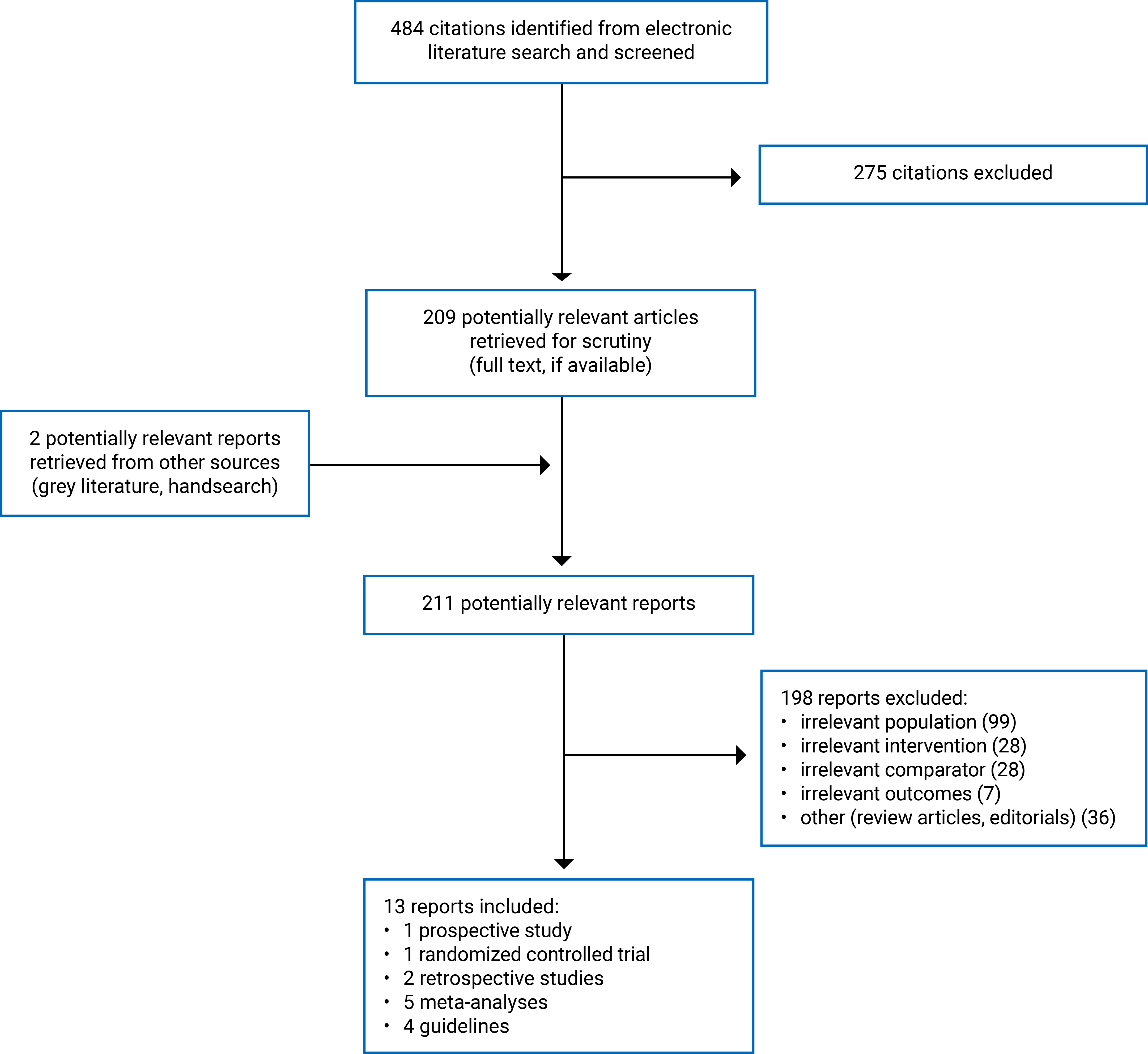
A total of 484 citations from the literature search were identified. Following screening of titles and abstracts, 275 citations were excluded, and 209 potentially relevant reports from the electronic search were retrieved for full-text review. Two potentially relevant publications from the grey literature search were also retrieved. Of these potentially relevant articles, 198 were excluded for various reasons. Overall, 13 publications met the inclusion criteria. These comprised 3 nonrandomized studies,24-26 2 of which were included from the previous report,25,261 randomized controlled trial (RCT),27 5 meta-analyses,13,28-31 and 4 evidence-based guidelines.32-35 The PRISMA36 flow chart of the study selection is available in Appendix 1, Figure 1.
Study Characteristics
Four primary studies and 5 meta-analyses were included in this review, totalling 11,041 patients across 8 countries who underwent CVC insertion using POCUS guidance or the landmark method.
Four evidence-based guidelines were included in this review to provide clinical guidance on the use of POCUS for CVC insertion in the emergency medicine, critical care, and acute care settings.
Study Design
Primary Studies and Meta-Analyses
Characteristics of the 9 included studies are presented in Appendix 2, Table 3:
Four primary studies24-27 (1 randomized controlled trial, 1 prospective non-RCT, 1 retrospective case-control study,1 retrospective cohort study) were published between 2020 and 2023.
Five meta-analyses were published between 2015 and 2024 and included 48 relevant primary studies published between 1989 and 2020 (46 RCTs, 1 prospective non-RCT, 1 retrospective non-RCT).13,28-31
The studies from each meta-analysis relevant to this report were included for analysis. The meta-analysis by Popat et al. (2024)30 included 81 publications, of which 2 were relevant to the present report. The meta-analyses by Zhou et al. (2022)31 and Gurien at al. (2018)13 included 5 and 3 RCTs, respectively. The meta-analysis by Brass et al. (2015a)28 included 35 publications, of which 29 were relevant to the present report. The meta-analysis by Brass et al. (2015b)29 included 13 studies, of which 9 were relevant to the present report. The meta-analyses did not include overlapping studies.
Evidence-Based Guidelines
Characteristics of the 4 evidence-based guidelines32-35 are presented in Appendix 2, Table 4. The 4 evidence-based guidelines include:
The European Society of Anesthesiology (ESA) guidelines by Lamperti et al. (2020):32
The authors conducted a systematic literature review from multiple databases and used the Grading of Recommendations Assessment, Development, and Evaluation (GRADE) tool to assess the level of evidence and strength of recommendations.
The European Society of Pediatric and Neonatal Intensive Care (ESPNIC) guidelines by Singh et al. (2020):33
The authors conducted a literature search for relevant studies from PubMed and used the GRADE tool.
The Society of Hospital Medicine (SHM) guidelines by Franco-Sadud et al. (2019):34
The authors conducted a systematic literature review from multiple databases. The quality of evidence and strength of recommendations were established by consensus using the Research and Development (RAND) Appropriateness method.
Society of Critical Care Medicine (SCCM) guidelines by Frankel et al. (2015):35
The authors conducted a systematic evidence search. The quality of evidence and strength of recommendations were established by consensus using GRADE and RAND, which included a modified Delphi method.
Country of Origin
The included primary studies were conducted by authors in India, Israel, the Republic of Korea, and Taiwan.24-27
The meta-analyses were conducted by authors in China, India, and the US.13,28-31 For 3 meta-analyses,2,20,27 the included studies were reported to have originated from 5 countries: Brazil, China, Italy, the Republic of Korea, and the US. Two meta-analyses28,29 did not report the country of origin for the studies included. All studies were conducted at a single institution (i.e., hospital).
The authors of the ESA and ESPNIC guidelines32,33 were reporting from Europe, while those of the SHM guidelines34 and SCCM guidelines35 were reporting from the US.
Patient Population
A summary of the patient populations and clinical settings are provided in Appendix 2, Table 3 for the primary studies and the systematic reviews.
The 4 primary studies24-27 included 1,903 adult and neonatal patients. In 2 studies,26,27 PICC was used to insert the CVC, and in 1 study27 UVC was used. Veins accessed for PICC insertion included: basilic, brachial, antecubital, and cephalic veins.
The 9,138 participants from the relevant studies included in the 5 meta-analyses were adults or children who were admitted to the ICU, emergency department, and nonemergency setting for central line insertion for various reasons. Veins accessed for CVC insertion included internal jugular,13,28,30 subclavian/axillary,13,29-31 and femoral veins.13,29-31
The target population in the ESA guidelines32 was adults and children who underwent vascular cannulation. The intended users were clinicians involved in perioperative procedures.
The target population in the ESPNIC guidelines33 was critically ill neonates and children in the pediatric ICU. The intended users were critical care providers and clinicians working in the pediatric ICU setting.
The target population in the SHM guidelines34 was acutely ill adult patients who required central venous access. The intended users were health care providers and clinicians who routinely place central and peripheral access catheters in acutely ill patients.
The target population in the SCCM guidelines35 were critically ill or injured adult patients who required central venous access. The intended users were health care providers and clinicians who routinely place central and peripheral access catheters in critically ill patients.
Interventions and Comparators
Appendix 5 details the terminology used to describe the intervention in this report and variations provided in the included studies.
The intervention used in all studies included in this report was POCUS guidance for CVC insertion to receive medications, fluids, nutrients, and other interventions.
The authors of the 4 included primary studies compared the following:
CVC inserted under POCUS guidance at a medical ICU versus CVC inserted using the landmark method at a surgical ICU.25 The authors also examined catheter wire localization
UVC inserted under POCUS guidance at a neonatal ICU versus UVC inserted using standard placement27
PICC inserted under POCUS or ultrasound guidance at bedside versus PICC inserted using blind or conventional methods (synonymous with the landmark method) at bedside.24,26
The authors of the 5 included meta-analyses compared the following:
CVC inserted under POCUS guidance versus CVC inserted using the landmark method in emergency departments30
CVC inserted under POCUS guidance via the axillary vein versus CVC inserted into the subclavian vein using the landmark method in the ICU setting31
Surgeon-preformed CVC insertion under real-time ultrasound guidance versus surgeon-performed CVC insertion using the landmark method in various nonemergency settings13
CVC inserted in the internal jugular vein under 2-dimensional ultrasound guidance versus CVC inserted using the landmark method in various hospital settings28
CVC inserted in the subclavian and femoral veins under 2-dimensional ultrasound guidance versus CVC inserted using the landmark method in various hospital settings;29 outcomes for the 2 veins were analyzed and reported separately.
Guidelines
Authors of the included guidelines considered the use of POCUS guidance for central venous line insertion in adult,32,34,35 pediatric, and neonatal patients.32,33
Outcomes
The relevant outcomes reported by the included studies are summarized in Table 2.
Table 2: Reported Outcomes in Included Studies
Type of outcome | Description |
|---|---|
Procedural characteristics | |
Complications |
The included guidelines32-35 considered all safety and effectiveness outcomes on the use of POCUS guidance for vascular access.
Summary of Findings
POCUS-guided CVC insertion resulted in a significantly higher correct placement rate compared to CVC insertion guided by the landmark method in most studies (92% versus 75%).
The use of POCUS-guided CVC insertion resulted in a decreased incidence of mispositioned catheters in 3 of 4 primary studies and 1 of 2 studies included in a meta-analysis.
The rate of technical failure was not significantly different between the POCUS-guided and landmark method groups, as reported by 3 primary studies and 1 study included in a meta-analysis.
POCUS-guided CVC resulted in significantly lower complication rates for pneumo- and hemothorax, arterial puncture, and hematoma compared to the landmark method when the internal jugular vein was targeted (0.9% versus 5.2%), whereas insertion at the femoral or subclavian veins resulted in variable findings.
All 4 guidelines provided recommendations for the use of POCUS-guided CVC insertion in emergency medicine, critical care, and acute care settings, although the strength of the recommendation varied depending on the targeted central vein.
Appendix 3 presents the main study findings.
Table 5 summarizes procedure-related outcomes (i.e., correct placement, malposition, technical failure, and procedure time).13,24-28,30,31
Table 6 summarizes outcomes related to complications (i.e., pneumothorax and hemothorax, arterial bleed, thrombosis, infection, hematoma, and overall complication rate).13,24-28,30,31
Table 7 summarizes the recommendations of the guidelines included as relevant to the acute care setting (e.g., emergency or intensive care).32-35
The primary studies and meta-analyses included in this report did not report on outcomes relating to mortality, hospital stay duration, and physician preference in CVC method of insertion.
Clinical Effectiveness of POCUS for Guided CVC Insertion
Procedural Characteristics
Figure 1: Summary of Statistical Findings by Procedure-Related Outcome
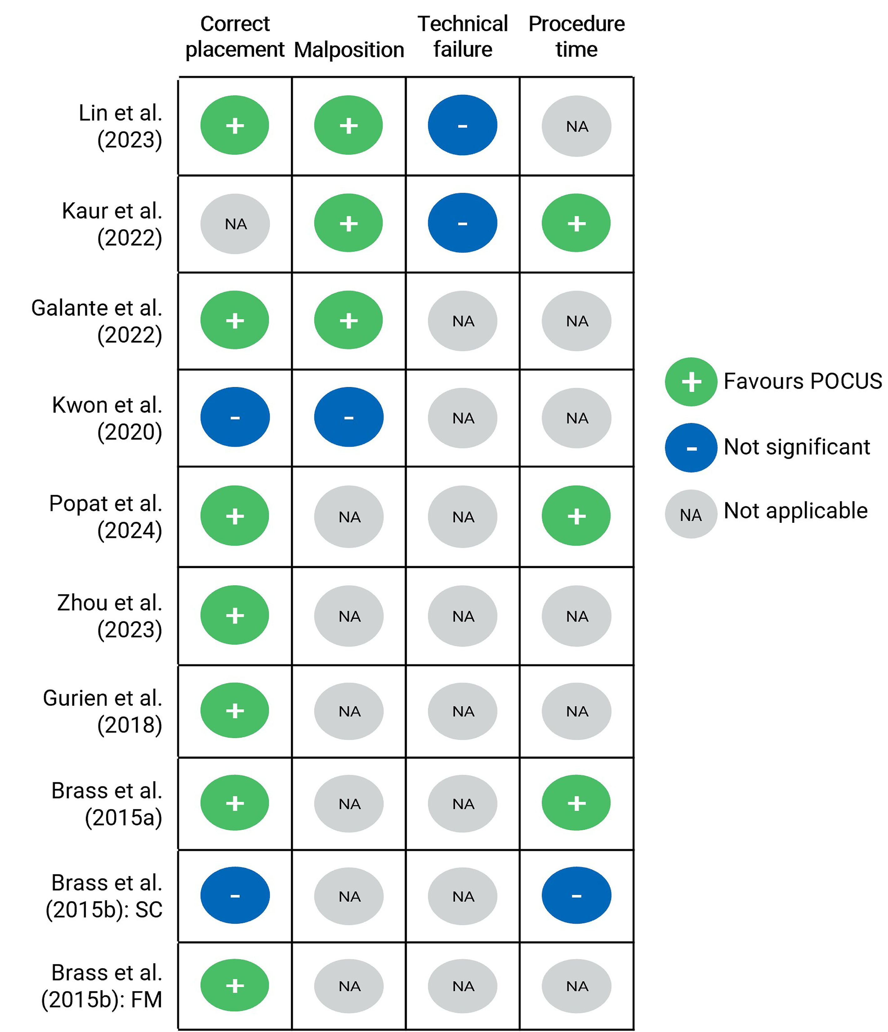
FM = femoral vein; POCUS = point-of-care ultrasound; NA = not applicable; SC = subclavian vein.
Note: The coloured circles represent the findings from studies that compared procedure-related outcomes between the POCUS and landmark method groups. Brass et al. (2015b)28 is represented by 2 rows to reflect the separate analyses conducted for the subclavian and femoral veins.
Green with a plus sign = POCUS guidance resulted in a statistically significant improved outcome compared to the landmark method (P < 0.05).
Blue with a minus sign = There was no statistically significant difference reported between groups (P ≥ 0.05).
Grey with NA = The study did not report on this outcome, no statistical comparisons were made, or in the case of the meta-analyses, no pooled analyses were conducted.
Correct Placement
Eight of the 9 studies reported this outcome.13,24-26,28-31
Two24,25 of the 4 primary studies reported that CVC insertion using POCUS resulted in a statistically significantly higher correct placement rate compared to CVC insertion by the landmark method.
Lin et al. (2023)24 reported that POCUS during PICC insertion at a neonatal ICU resulted in a statistically significantly higher correct placement rate compared to that of CVC insertion by the conventional method (87.3% versus 31.3%; P < 0.001).
Galante et al. (2022)25 reported that CVC insertion using POCUS for guidewire visualization in a medical ICU resulted in a statistically significantly higher correct placement rate compared to that of CVC insertion by traditional method in a surgical ICU (97.6% versus 88.0%; P = 0.001).
Kwon et al. (2020)26 compared POCUS-guided PICC insertion to PICC insertion using the landmark method and reported no statistically significant difference in correct placement rates (98.6% versus 97.1%; P = 0.05).
Kaur et al. (2022)27 did not investigate correct placement rates between POCUS and landmark method groups.
Four13,28,30,31 of the 5 meta-analyses reported that CVC insertion using POCUS resulted in a statistically significantly higher correct placement rate compared to that of CVC insertion by the landmark method. Bass et al. (2015b)29 reported mixed findings.
The meta-analysis by Popat et al. (2024)30 included 2 studies that compared POCUS-guided CVC insertion to CVC insertion using the landmark method in the emergency department. In 1 study, the correct placement rate was significantly higher when using POCUS compared to CVC insertion without POCUS (98% versus 79%; OR = 13.1; 95% confidence interval [CI], 2.0 to 59.4). The second study used the average time to successful insertion as an indirect measure of correct placement. Popat et al. reported that POCUS resulted in a reduced time to successful placement compared with the landmark method (P < 0.0001).
Zhou et al. (2023)31 conducted a pooled analysis of 5 studies that examined POCUS-guided CVC insertion via the axillary vein in the ICU setting. The authors reported that the POCUS group showed significantly higher rates of successful placement compared to the landmark method group (98.3% versus 89.1%, RR = 1.09; 95% CI, 1.04 to 1.15; P < 0.001).
Similarly, Gurien et al. (2018)13 conducted a pooled analysis of 3 studies that examined POCUS-guided CVC insertion in the nonemergency setting. The authors reported that the POCUS approach resulted in a statistically significantly higher rate of successful placements compared to the landmark method (97.1% versus 83.3%; P < 0.0001).
Bass et al. (2015a)28 conducted a pooled analysis of 23 studies that examined correct placement using 2-dimensional ultrasound-guided CVC insertion via the internal jugular vein. The authors reported that the ultrasound group showed significantly higher rates of successful placement compared to the landmark method group, for both first attempts (79.7% versus 50.1%) and overall attempts (97.6% versus 87.6%; P < 0.0001).
Bass et al. (2015b)29 conducted a pooled analysis of 3 studies that examined correct placement using two-dimensional ultrasound-guided CVC insertion via the subclavian vein. The authors reported no statistically significant differences in the success rates of first and overall attempts between the ultrasound and landmark method groups (P > 0.1).
Bass et al. (2015b)29 also conducted a pooled analysis of 4 studies that examined correct placement using 2-dimensional ultrasound-guided CVC insertion via the femoral vein. The authors reported that the ultrasound group showed a statistically significantly higher rate of success during first attempts compared to the landmark method group (85% versus 48.7%; P < 0.0001), although the overall success rates did not significantly differ between groups (P = 0.06).
Malposition
This outcome was reported by all primary studies.24-27 Malposition was not included in the pooled analysis for any of the meta-analyses,13,28-31 although 2 studies analyzed by Zhou et al. (2022)31 did report on this outcome. The results of this outcome were mixed, following a similar pattern to the results for correct placement:
Three primary articles24,25,27 reported that the rate of CVC tip malposition was significantly lower for POCUS-guided CVC insertion compared to landmark method–guided CVC insertion (12.7% versus 62.5%; P < 0.001),24 (2.4% versus 12%; P = 0.001),25 (42.3% versus 74%; P = 0.019).27
Kwon et al. (2020)26 reported a non–statistically significant difference in the rates of CVC tip malposition for patients in the ultrasound-guided group compared with patients in the landmark method group (9.8% versus 5.8%; P = 0.152).
Two studies included in the meta-analysis conducted by Zhou et al. (2022)31 reported variable findings. While 1 RCT reported a significantly lower CVC tip malposition rate in the POCUS group compared to the landmark method group (0.6% versus 2.1%; P = 0.017), another RCT reported no statistically significant difference between the 2 groups (4% versus 13%; P = 0.331).
Technical Failure
Technical failure was reported by 3 primary studies,24,26,27 although only 2 studies made statistical comparisons.24,27 Technical failure was not included in the pooled analysis for any of the meta-analyses, although 3 studies reviewed by Gurien et al. (2018)13 and 1 study analyzed by Zhou et al. (2022)31 included technical failure as an outcome. Generally, there were no significant differences in the rates of CVC technical failures between POCUS-guided and landmark method groups.
For all 7 studies that reported technical failure, a lower failure rate was reported for the POCUS-guided insertion group compared to the landmark method group, although statistical significance was reached in only 1 study (2.6% versus 22.2%; P = 0.009).13
Procedure Time
Procedure time was reported as an outcome by 2 primary studies24,27 and was included in the pooled analysis for 2 meta-analyses.28,29 Overall, the results were mixed:
The primary study by Kaur et al. (2022) conducted statistical comparisons and found that the procedure time was 22% faster on average for the POCUS-guided group compared to the landmark method group (P = 0.005).27
The primary study by Lin et al. (2023)24 reported procedure time for the POCUS-guided group (IQR 360 to 900 seconds), but not the landmark method group. No statistical comparisons were reported for this outcome.
Procedure time was included in the pooled analysis for 228,29 of the 5 meta-analyses:
Brass et al. (2015a)28 reported that when the internal jugular vein was targeted, ultrasound guidance decreased the time (−30.5 seconds) to successful CVC insertion compared to the use of the landmark method (P = 0.02).
Brass et al. (2015b)29 reported that when the subclavian vein was targeted, no statistically significant differences were observed between groups (P = 0.76). The authors did not report on this outcome when targeting the femoral vein due to the lack of available data.
Procedure time was not included in the pooled analysis for 3 meta-analyses,13,30,31 although 7 studies reviewed as part of the meta-analyses reported this outcome:13,30,31
The 7 relevant studies reported that POCUS guidance decreased the average time to successful CVC insertion compared to the use of the landmark method.
Four of the 7 relevant studies within the meta-analyses performed statistical comparisons:
Complications
Three primary studies24,25,27 and all 5 meta-analyses13,28-31 reported complications associated with CVC insertion guided by POCUS and the landmark method. All 5 meta-analyses made statistical comparisons, whereas most primary studies did not. Overall, the findings were variable and are based on limited data that are available for complication-related outcomes.
Results from the meta-analyses indicated that complication rates were significantly lower in the POCUS group compared with the landmark method groups for the internal jugular and axillary veins, but results were inconsistent for the subclavian and femoral veins. Figure 2 presents a summary of statistical findings by study.
Figure 2: Summary of Statistical Findings by Complication
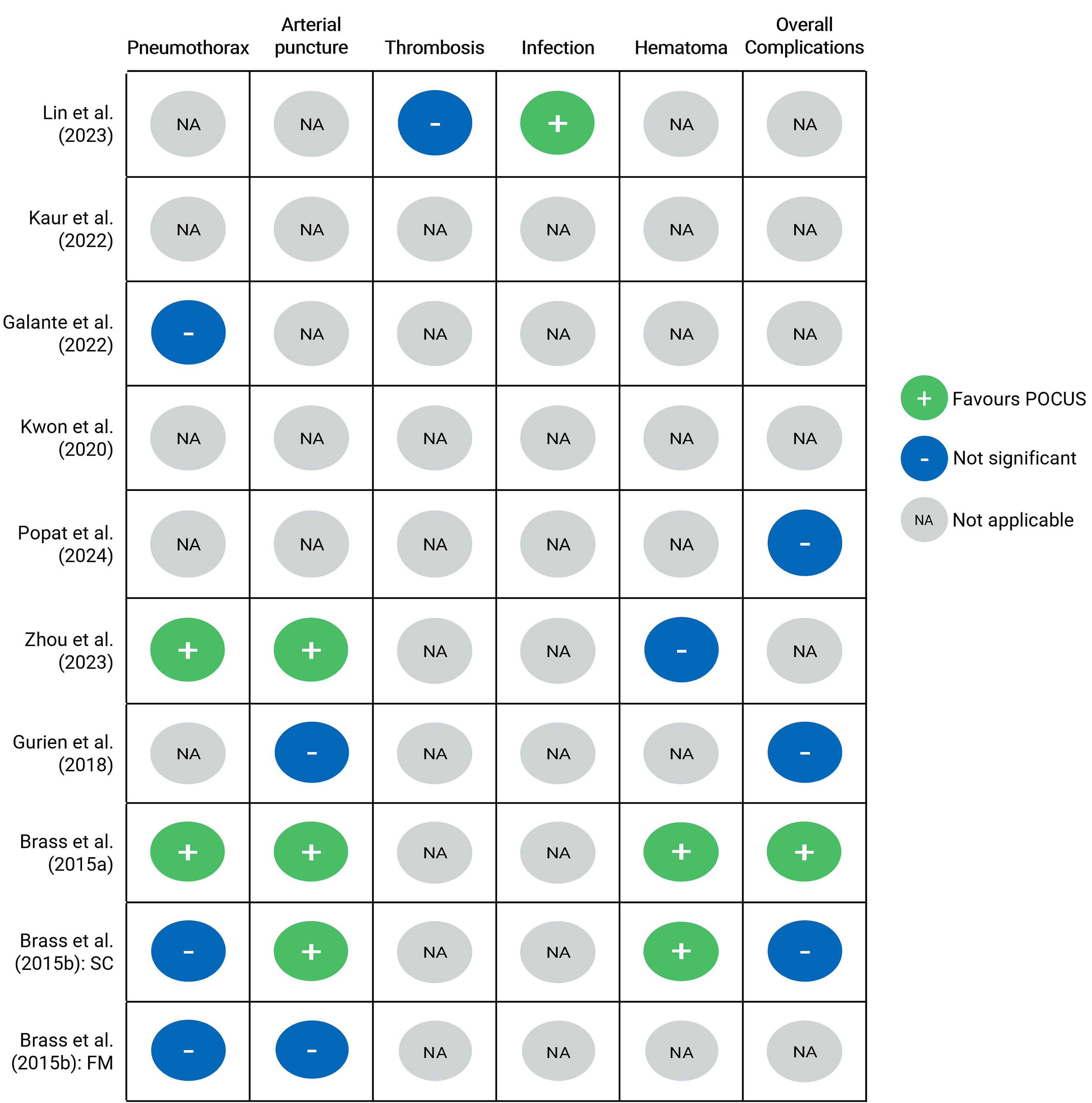
FM = femoral vein; NA = not applicable; POCUS = point-of-care ultrasound; SC = subclavian vein
Note: The coloured circles represent the findings from studies that compared complication-related outcomes between the POCUS and landmark method groups. Brass et al. (2015b)29 is represented by 2 rows to reflect the separate analyses conducted for the subclavian and femoral veins.
Green with a plus sign = POCUS guidance resulted in a statistically significant improved outcome compared to the landmark method.
Blue with a minus sign = There were no reported statistically significant differences between groups (P ≥ 0.05).
Grey with NA = The study did not report on this outcome, no statistical comparisons were made, or in the case of the meta-analyses, no pooled analysis was conducted.
Pneumothorax or Hemothorax
The results were mixed for this complication. One primary study25 and 3 meta-analyses31 conducted statistical testing to compare the incidence of pneumothorax or hemothorax between POCUS-guided CVC insertion and LM-guided CVC insertion.
The 2 meta-analyses conducted by Brass et al. (2015a)28 and Brass et al. (2015b)29 included a composite outcome that captured various kinds of complications, including pneumothorax and hemothorax, embolism, thrombosis, and others. In this report, this composite outcome is reported in this section.
In the pooled analysis of 5 studies that was conducted by Zhou et al. (2022),31 the authors noted a statistically significant difference in pneumothorax or hemothorax rates (0% versus 1.2%; RR = 0.12; 95% CI, 0.02 to 0.64; P = 0.01), whereas Galante et al. (2022)25 did not find a statistically significant difference among groups (P > 0.05).
In the pooled analysis of 11 studies with data on pneumo- or hemothorax that examined CVC insertion at the internal jugular vein, Brass et al. (2015a)28 noted a statistically significant difference in the rate of pneumothorax or hemothorax (composite outcome: 0.6% versus 3%; P = 0.01).
In the 2 pooled analyses that examined CVC insertion at the subclavian vein (3 studies) and femoral vein (4 studies), Brass et al. (2015b)29 did not report a statistically significant difference among POCUS-guided and landmark method groups for both the subclavian and femoral veins (subclavian vein composite outcome: 0.8% versus 13.3%; P = 0.31; femoral vein composite outcome: 1.3% versus 3.1%; P = 0.34).
Arterial Puncture
Results for arterial puncture complications were also mixed. Four meta-analyses13,28,29,31 conducted 5 pooled analyses for arterial puncture incidence following CVC insertion.28,29 This outcome was not reported in any of the primary studies.
Three of the 5 pooled analyses showed statistically significantly lower arterial puncture rates in the POCUS-guided CVC group (0.2% to 2.0%) compared to the landmark method group (2.1% to 9.4%) when the subclavian29,31 or internal jugular28 veins were targeted.
Gurien et al. (2018)13 reported no significant difference in arterial puncture rates between the POCUS-guided and landmark method groups when the subclavian and internal jugular veins were targeted (P = 0.18). Similarly, Brass et al. (2015b)29 did not find a statistically significant difference in puncture rates when the femoral vein was targeted (P = 0.09).
Thrombosis
One primary study24 compared thrombosis rates between POCUS-guided PICC insertion and PICC insertion using the landmark method.
There was no statistically significant difference in thrombosis rates between the 2 groups (0% versus 6%, P = 0.319).
Brass et al. (2015a)28 and Brass et al. (2015b)29 reported on thrombosis as part of a composite outcome that is presented in the “Pneumothorax or Hemothorax” section of this report.
Infection
One primary study compared the rate of infections between POCUS-guided PICC insertion and landmark method–guided PICC insertion.24
Three types of infections were analyzed: CLABSI, catheter-related bloodstream infection (CRBSI), and sepsis. The POCUS-guided group experienced a statistically significantly lower rate of CRBSI (0% versus 6.3%; P = 0.044) than the landmark method group. There were no statistically significant differences in the rates of CLABSI or sepsis between the POCUS and landmark method groups.
Hematoma
Three meta-analyses statistically compared hematoma occurrence between POCUS-guided CVC insertion and CVC inserted using the landmark method.28,29,31
Two28,29 of the 3 meta-analyses reported statistically significantly lower hematoma rates for the POCUS-guided group (1.2% to 1.6%) compared to the landmark method group (6.6% to 6.7%), whereas the other meta-analysis31 found no statistically significant difference in hematoma rates between groups (P = 0.07).
Overall Complications
Four meta-analyses statistically compared the overall complication rate between POCUS-guided CVC insertion and CVC inserted using the landmark method.28,29,31
One28 of the 4 meta-analyses reported a statistically significantly lower rate of overall complications for the POCUS-guided group compared to the landmark method group (4.0 versus 13.5; P < 0.0001), while the other 3 meta-analyses13,29,30 reported no statistically significant difference in overall complication rates between groups (P > 0.1).
Evidence-Based Guidelines Regarding the Use of POCUS for Guided CVC Insertion
Various methods were used by each guideline to assess the quality and strength of the recommendations and to determine consensus on the use of POCUS-guided CVC insertion.
The ESA Guidelines32 on Perioperative Use of POCUS for Guided Vascular Access
POCUS-guided vascular cannulation in adults:
The ESA guideline provides recommendations for ultrasound-guided (including POCUS-guided) cannulation in adults via the internal jugular vein, the subclavian vein, the axillary vein, the femoral vein, the peripheral vein, and any central vein for long-term vascular access device placement.
The ESA guideline makes strong recommendations on the use of ultrasound guidance for internal jugular vein cannulation, for subclavian vein cannulation, for femoral vein cannulation, and for peripheral vein cannulation. Low- to moderate-quality evidence supported these recommendations.
The ESA guideline makes a weak recommendation on the use of ultrasound guidance for axillary vein cannulation, based on low-quality evidence.
The ESA guideline makes strong recommendations to use ultrasound guidance for placement of long-term vascular access devices via the axillary vein at the thorax, for catheter tip location and tip navigation, for preprocedural sonographic evaluation of all possible venous option, and for timely diagnosis of all potentially life-threatening complications (pneumothorax, hemothorax, cardiac tamponade, and so forth) after central venipuncture. Low- to moderate-quality evidence supported these recommendations.
POCUS-guided vascular cannulation in children:
The ESA guideline provides recommendations for POCUS-guided cannulation in children via the internal jugular vein, the brachiocephalic vein, the femoral vein, and the peripheral vein.
The ESA guideline makes strong recommendations on the use of ultrasound guidance for internal jugular vein cannulation, for brachiocephalic vein cannulation, and for femoral cannulation. Low- to moderate-quality evidence supported these recommendations.
The ESA guideline makes a weak recommendation for routine use of ultrasound guidance for peripheral vein cannulation, based on moderate-quality evidence.
The ESPNIC Guideline33 on the Use of POCUS in Critically Ill Neonates and Children
For neonates and children, the ESPNIC guideline makes strong recommendations on the use of the POCUS-guided technique to place central lines in the internal jugular vein, the subclavian vein, and the femoral vein. Supporting evidence for these recommendations was graded as level A or B, reflecting high or moderate confidence in the estimate of effect, respectively.
For children, the ESPNIC guideline makes a moderate recommendation for the use of the POCUS-guided technique for PICC insertion, based on evidence with a quality level of B.
The SHM Guideline34 on the use of Ultrasound Guidance for Central and Peripheral Vascular Access in Adults
The SHM guideline provides recommendations on central venous access techniques and peripheral venous access techniques, including “real-time ultrasound guidance” (i.e., POCUS guidance).
The SHM guideline makes strong recommendations, with very good consensus, on the use of real-time ultrasound guidance for internal jugular vein catheterization, for subclavian vein catheterization, and for femoral venous access.
The SHM guideline also makes a strong recommendation, with very good consensus, on the use of real-time ultrasound guidance for the insertion of PICC.
The SCCM Guideline35 on the Use of Bedside General and Cardiac Ultrasound Guidance for Central Vascular Access in Adult Patients
The SCCM guideline makes strong recommendations, with very good consensus, on the use of ultrasound guidance for internal jugular vein catheterization to improve success rate, shorten procedure time, and reduce the risk of procedure-related complications. The quality of supporting evidence was graded as A or “high.”
The SCCM guideline suggests that real-time ultrasound guidance for subclavian vein catheterization is of limited value in adult patients. The quality of supporting evidence was graded as C or “low” and the strength of the recommendation was conditional (weak).
The SCCM guideline makes strong recommendations, with very good consensus, on the use of real-time ultrasound guidance for femoral vein catheterization to improve success rate and reduce complications in adults, although this benefit is most relevant to novice operators. The quality of supporting evidence was graded as A or “high.”
The SCCM guideline makes strong recommendations, with very good consensus, that in most patients, real-time ultrasound is preferred over static preprocedure marking. The quality of supporting evidence was graded as B or “moderate.”
We did not identify any Canadian evidence-based guidelines regarding the use of POCUS guidance for CVC insertion, although a consensus statement was released by the Canadian Internal Medicine Ultrasound group on recommended POCUS competencies for ultrasound-guided CVC insertion.37
Summary of Critical Appraisal
Appendix 4 provides details regarding the strengths and limitations of the included primary studies (Table 8),24-27 meta-analyses (Table 9),13,28-31 and guidelines (Table 10).32-35
Primary Studies
The included studies24-27 were explicit in terms of reporting but had several limitations regarding their external and internal validity that may reduce the certainty and generalizability of the findings.
For reporting, the authors of all included studies24-27 clearly described the objective of the study, main outcomes to be measured, characteristics of the participants included in the study, interventions of interest, and main findings. The authors reported actual P values for the main outcomes in all studies.24-27 The authors of 3 studies24,25,27 reported adverse events of the intervention.
For the external validity of the treatment settings (i.e., hospitals), all studies24-27 were representative of the treatment typically received by most patients. However, the patient populations may not be representative of the broader population from which they were selected, potentially limiting the generalizability of the findings to different settings or patient groups outside of the studies; 2 of the 4 primary studies and 1 meta-analysis included retrospective chart review data.25,26,30 Additionally, there is some uncertainty around how generalizable these results are within the Canadian context.
For internal validity related to bias, 2 studies25,26 were of retrospective design, which introduces several limitations, including risks of selection bias and missing data. However, the authors used statistical tests appropriately for comparing variables and assessed the main outcomes using accurate and reliable methods (i.e., chest X-ray) to verify catheter tip position and the presence of potential complications immediately after catheter insertion. The nonrandomized study24 had a selection bias due to limited availability of POCUS equipment and trained operators, which may have introduced differences between groups that impacted statistical comparisons.
For internal validity related to confounding, there were some differences between patient groups in demographics and clinical features, as well as in the experience of operators who performed the CVC insertion procedures. The authors of all included studies did not identify and adjust for potential confounding factors in the analyses. For instance, in 1 study,25 patients in the POCUS group frequently presented more severely and with more underlying comorbidities compared to those in the control groups. Authors in 2 of the primary studies did not report whether sample size calculations were performed, and it is unclear whether the nonsignificant differences in certain outcomes were because the studies were underpowered to accurately measure those outcomes.
Meta-Analyses
Overall, the 5 meta-analyses13,28-31 met most of the AMSTAR criteria, indicating a high level of methodological rigour. The reviews were comprehensive in their search strategies, clearly defined their inclusion criteria, and were thorough in their assessment of study quality and heterogeneity. The authors of 3 meta-analyses13,30,31 reported low or no heterogeneity among the included studies, while 2 authors28,29 noted high heterogeneity for several outcomes.
The authors of all 5 meta-analyses13,28-31 included components of the PICO that were clearly defined in research questions and inclusion criteria. All authors explained their selection of eligible study designs, which were either nonrandomized and randomized studies30 or randomized studies alone.13,31 All authors conducted a comprehensive literature search using multiple databases and a review of bibliographies for potentially relevant studies.13,28-31 All review authors performed study selection, extraction, and quality assessment in duplicate.13,28-31
The review authors used a satisfactory technique for assessing the quality of the included studies. The QUADAS-2 checklist and Cochrane Risk-of-Bias Tool were used in 1 report,30 the Cochrane Risk-of-Bias Tool was used alone in 2 reports,13,31 and the domain-based evaluation table of the Cochrane Collaboration was used in 2 reports.28,29 It is noted by the review authors in 4 reports13,28,29,31 that CVC insertion operators were not blinded in the included studies, which introduces potential bias. One report30 included nonrandomized studies, which introduces selection and confounding bias to favour POCUS. All review authors disclosed the funding sources and potential conflicts of interest for the included studies.13,28-31
Two28,29 of the 5 meta-analyses included a list of excluded studies and reasons for study exclusion. None of the meta-analyses reported the funding sources for the included studies.13,28-31
Evidence-Based Guidelines
The included evidence-based guidelines32-35 had several strengths related to reporting. They were explicit in terms of scope and purpose (i.e., objectives, health questions, and populations) and provided clear presentations of recommendations (i.e., specific, unambiguous, and easy-to-find key recommendations with options for managing different conditions or health issues).
In terms of the involvement of interested parties, the authors of all included guidelines32-35 clearly defined target users and the development groups but did not report whether the views and preferences of patients were sought. The methodology for the development of all included guidelines32-35 was robust. The authors of the guidelines32-35 clearly reported methods for evidence collection, criteria for selection, and methods for evidence synthesis. There were explicit links between the recommendations made and the supporting evidence and methods of formulating the recommendations in all guidelines.32-35 Also, the authors of all guidelines32-35 considered health benefits and risks of side effects when formulating their recommendations.
However, there were also some limitations related to guideline implementation and review. Specifically, the authors did not report on the procedures for updating the guidelines.32-35
Additionally, for 1 of the guidelines,35 the specific health questions covered were not clearly described. Facilitators and barriers to application, advice and/or tools on how the recommendations can be put into practice, and monitoring or auditing criteria were also unclear in the included guidelines.32-35 For editorial independence, the authors of 3 guidelines32-34 reported that all guideline development group members had no competing interests. However, the authors of 2 guidelines32,34 did not report if the views of the funding body had any influence on the content of the guidelines.
Some recommendations were developed primarily on generally weak evidence, with studies having a high degree of heterogeneity due to different patient populations, settings, and operators performing the procedures. As ultrasound guidance is not a therapeutic intervention but rather a diagnostic and monitoring technique, evidence supporting the recommendations was mostly derived from short-term outcomes.
Overall, all 4 included guidelines32-35 were robust in terms of scope and development, rigour of development, and clarity of presentation, although their generalizability to the Canadian context remains unclear.
Limitations
This report is limited by the quantity and quality of research identified that met our inclusion criteria. First, the primary studies and systematic reviews we identified are at risk of bias due to several important limitations outlined in the critical appraisal section.
Second, the literature search was limited to English-language articles and primary articles published within the past 5 years and systematic reviews published after 2013. While the studies mentioned in this report include a total of more than 11,000 participants across a 35-year span, the use of POCUS for CVC insertion has been investigated before 2013. Therefore, the strength of the conclusions in this report may be limited by the exclusion of relevant articles published before 2013.
Third, patients who underwent POCUS-guided CVC at the bedside usually were critically ill and had a higher rate of underlying comorbidities compared to patients who did not undergo CVC insertion at the bedside, who were relatively healthier. Both patient groups had different causes of ICU admission. POCUS-guided bedside insertion would be difficult to apply in patients who had been hospitalized with prolonged chronic disease, as they tend to have poor venous access. It is, therefore, possible that the success rate of the CVC insertion procedure may vary depending on the skill of the operator and the medical complexity of the patient undergoing the procedure.
Fourth, all included studies reported short-term in-hospital clinical outcomes during admission, such as correct placement of central line and procedural complications that occurred right after catheter insertion. However, the included studies did not investigate late complications over a longer duration of use, such as central vein stenosis and complication-related mortality. In general, due to differences in patients’ characteristics and operators’ competence between groups, it is hard to conclude whether POCUS-guided central line insertion is effective compared to the landmark method. The evidence found in this report appeared to be institution-specific, and therefore, it may not be generalizable to other institutions.
Conclusions
We reviewed the clinical evidence from 4 primary studies24-27 (1 RCT, 1 nonrandomized study, 1 retrospective case-control study, 1 retrospective cohort study) and 5 meta-analyses,13,28-31 all comparing POCUS-guided insertion of central lines at the bedside or in the surgical setting with the traditional landmark method using different vein locations.
The 4 primary studies identified in this report showed moderately high-quality evidence, although all were limited by a single-centre study design and had limitations that may have reduced the generalizability and validity of findings. Similarly, the 5 meta-analyses showed high methodological rigour and included predominantly RCT studies.
Overall, the use of POCUS to guide the insertion of CVC placement is effective compared with the landmark method. All publications with data, apart from 1, reported that CVC insertion using POCUS resulted in a statistically significantly higher correct placement rate compared to that of CVC insertion by the landmark method. POCUS guidance for central line insertion at the bedside appeared to be safe with few complications (e.g., pneumothorax, bleeding, infection) compared to the use of the landmark method.
The differences in patients’ characteristics, operators’ performance, which vein was targeted for CVC insertion, and how the procedure was performed may contribute to the mixed findings in success rates and certain outcomes between groups (e.g., insertion duration and physician preferences in the CVC method of insertion).
Four evidence-based guidelines32-35 provided detailed recommendations on the use of POCUS-guided vascular access in children and adults in emergency medicine, critical care, and acute care settings. Overall, the evidence that informed the guidelines ranged from low to high quality.
All guidelines identified in this report support the use of ultrasound-guided cannulation using different vein locations in both adults and children. The strengths of the recommendations were generally strong despite mixed levels of evidence due to heterogeneity from previous RCTs and observational studies. However, 1 guideline suggested that the use of POCUS to guide CVC insertion at the subclavian vein is of limited value, but this was based on low-quality evidence. The publication of new literature may prompt a revision of the guidelines. Because the included guidelines were developed by authors in Europe and the US, the recommendations of the guidelines can likely be applied to clinical practice in Canada.
Implications for Decision- or Policy-Making
CVC insertion guided by anatomical landmarks may be unsuccessful in one-third of cases and is associated with a high rate of mechanical complications.28,29 In addition, anatomical variations in the size and location of the central veins have been described, which can make it difficult or impossible to perform the procedure without serious risk to the patient.
Conversely, POCUS-guided CVC allows for the direct visualization of the needle tip and target vessel, which can be effective and safe for short-term use, provided that the procedure is performed by a well-trained and experienced operator. POCUS devices are also more portable, less expensive, and easier to use than traditional diagnostic ultrasound.20
POCUS has gained widespread use in diverse clinical settings and been incorporated into bedside practice by an increasing number of health care professionals with limited or no training in imaging.38 Updated guidelines have been recently published by the SCCM, which recommends the use of POCUS to guide the management of several clinical conditions in the critical care setting.39
Future well-controlled studies are needed to evaluate both short-term and long-term outcomes of ultrasound guidance for central line insertion at the bedside. Because the use of ultrasound is considered the preferred method for CVC insertion by multiple organizations and societies, there is growing interest to understand how ultrasound may be most effectively used to support CVC insertion and promote patient outcomes.37 Appendix 6 provides a list of potential areas of interest that future studies could consider when determining how ultrasound will be used for CVC insertion.
In addition to guiding CVC insertion, POCUS may also play a role in confirming the correct placement of the catheter, which is typically achieved with X-ray by experienced professionals. The potential for POCUS to replace X-ray in this role can lower the use of hospital resources, reduce radiation exposure, and decrease the time delay to catheter use.
References
1.Tran K, Horton J. CADTH Health Technology Review: Point-of-care ultrasound for guided central venous catheter insertion. Can J Health Technol. 2023;3(10). Accessed 2025 Jan 6. https://www.cda-amc.ca/sites/default/files/pdf/htis/2023/RC1509-POCUS%20for%20CVC.pdf PubMed
2.Central venous catheter-associated blood stream infections in intensive care units in Canadian acute-care hospitals: Surveillance report January 1, 2006 to December 31, 2006 and January 1, 2009 to December 31, 2011. Public Health Agency of Canada; 2014. Accessed 2025 Jan 6. https://publications.gc.ca/collections/collection_2014/aspc-phac/HP40-90-2013-eng.pdf
3.Haddadin Y, Annamaraju P, Regunath H. Central line-associated blood stream infections. StatPearls Publishing; 2024. Accessed 2025 Jan 6.
4.Akmal AH, Hasan M, Mariam A. The incidence of complications of central venous catheters at an intensive care unit. Ann Thorac Med. 2007;2(2):61-3. doi:10.4103/1817-1737.32232 PubMed
5.Wong AV, Arora N, Olusanya O, et al. Insertion rates and complications of central lines in the UK population: A pilot study. J Intensive Care Soc. 2018;19(1):19-25. doi:10.1177/1751143717722914 PubMed
6.Tse A, Schick MA. Central line placement. StatPearls Publishing LLC.; 2024. Accessed 2025 Jan 6. https://www.ncbi.nlm.nih.gov/pubmed/29262231
7.Saint Luke's. Central line (central venous access device). Accessed 2025 Jan 6, https://www.saintlukeskc.org/health-library/central-line-central-venous-access-device
8.Heffner AC, Andros MP. Post TW, ed. Central venous access in adults: General principles of placement. UpToDate; 2023. Accessed 2025 Jan 6. https://www.uptodate.com
9.Chopra V. Post TW, ed. Central venous access: Device and site selection in adults. UpToDate; 2023. Accessed 2023 Sep 19. https://www.uptodate.com
10.Lewis K, Spirnak PW. Umbilical vein catheterization. StatPearls Publishing; 2023. Accessed 2024 Sep 3. https://www.ncbi.nlm.nih.gov/books/NBK549869/
11.Central venous catheter-related deep vein thrombosis. Thrombosis Canada; 2021. Accessed 2025 Jan 6. https://thrombosiscanada.ca/wp-uploads/uploads/2021/09/1.-Central-Venous-Catheter_14Sept2021.pdf
12.Bonz J, Wong A, Evans L. Implementation of a hospital mandated central venous catheter (CVC) training program. Ann Vasc Med Res. 2017;4(6):1074. doi:10.47739/2378-9344/1074 Accessed 2025 Jan 6. https://www.jscimedcentral.com/public/assets/articles/vascularmedicine-4-1074.pdf
13.Gurien LA, Blakely ML, Crandall MC, et al. Meta-analysis of surgeon-performed central line placement: Real-time ultrasound versus landmark technique. J Trauma Acute Care Surg. 2018;84(4):655-663. doi:10.1097/TA.0000000000001784 PubMed
14.Young MP. Post TW, ed. Central venous catheters: Overview of complications and prevention in adults. UpToDate; 2023. Accessed 2025 Jan 6. https://www.uptodate.com
15.Teja B, Bosch NA, Diep C, et al. Complication rates of central venous catheters: A systematic review and meta-analysis. Systematic Review Meta-Analysis Research Support, Non-U.S. Gov't Comment. JAMA Intern Med. 2024;184(5):474-482. doi:10.1001/jamainternmed.2023.8232 PubMed
16.Deere M, Singh A, Burns B. Central venous access of the subclavian vein. StatPearls Publishing; 2023. Accessed 2025 Jan 6. https://www.ncbi.nlm.nih.gov/books/NBK482224/
17.Bailey PL, Whitaker EE, Palmer LS, Glance LG. The accuracy of the central landmark used for central venous catheterization of the internal jugular vein. Anesth Analg. 2006;102(5):1327-32. doi:10.1213/01.ane.0000202467.10465.12 PubMed
18.Saugel B, Scheeren TWL, Teboul JL. Ultrasound-guided central venous catheter placement: A structured review and recommendations for clinical practice. Crit Care. 2017;21(1):225. doi:10.1186/s13054-017-1814-y PubMed
19.Moore CL, Copel JA. Point-of-care ultrasonography. N Engl J Med. 2011;364(8):749-57. doi:10.1056/NEJMra0909487 PubMed
20.Required and preferred ultrasound scanner features for conventional and point-of-care applications (updated 3/30/2022). ECRI; 2015.
21.Downs SH, Black N. The feasibility of creating a checklist for the assessment of the methodological quality both of randomised and non-randomised studies of health care interventions. J Epidemiol Community Health. 1998;52(6):377-384. doi:10.1136/jech.52.6.377 PubMed
22.Shea BJ, Reeves BC, Wells G, et al. AMSTAR 2: A critical appraisal tool for systematic reviews that include randomised or non-randomised studies of healthcare interventions, or both. BMJ. 2017;358:j4008. NOT IN FILE. doi:10.1136/bmj.j4008
23.Agree Next Steps Consortium. The AGREE II Instrument. AGREE Enterprise; 2017. Accessed 2025 Jan 6. https://www.agreetrust.org/wp-content/uploads/2017/12/AGREE-II-Users-Manual-and-23-item-Instrument-2009-Update-2017.pdf
24.Lin SY, Chiang MC, Wu WH, et al. Point-of-care ultrasound (POCUS) for tip localization of neonatal peripherally inserted central catheter (PICC): A prospective study. Pediatr neonatol. 2024;65(4):375-380. doi:10.1016/j.pedneo.2023.07.008 PubMed
25.Galante O, Sadeh R, Smoliakov A, et al. Real time wire localization by ultrasound during central line insertion improves accurate positioning in all upper torso sites. J Crit Care. 2022;67:79-84. doi:10.1016/j.jcrc.2021.10.009 PubMed
26.Kwon S, Son SM, Lee SH, et al. Outcomes of bedside peripherally inserted central catheter placement: A retrospective study at a single institution. Acute Crit Care. 2020;35(1):31-37. doi:10.4266/acc.2019.00731 PubMed
27.Kaur A, Manerkar S, Patra S, Kalamdani P, Kalathingal T, Mondkar J. Ultrasound-guided umbilical venous catheter insertion to reduce rate of catheter tip malposition in neonates: A randomized, controlled trial. Randomized Controlled Trial. Indian J Pediatr. 2022;89(11):1093-1098. doi:10.1007/s12098-022-04295-w PubMed
28.Brass P, Hellmich M, Kolodziej L, Schick G, Smith AF. Ultrasound guidance versus anatomical landmarks for internal jugular vein catheterization. Cochrane Database Syst Rev. 2015;1(1):CD006962. doi:10.1002/14651858.CD006962.pub2 PubMed
29.Brass P, Hellmich M, Kolodziej L, Schick G, Smith AF. Ultrasound guidance versus anatomical landmarks for subclavian or femoral vein catheterization. Cochrane Database Syst Rev. 2015;1(1):CD011447. doi:10.1002/14651858.CD011447 PubMed
30.Popat A, Harikrishnan S, Seby N, et al. Utilization of point-of-care ultrasound as an imaging modality in the emergency department: A systematic review and meta-analysis. Review. Cureus. 2024;16(1):e52371. doi:10.7759/cureus.52371 PubMed
31.Zhou J, Wu L, Zhang C, Wang J, Liu Y, Ping L. Ultrasound guided axillary vein catheterization versus subclavian vein cannulation with landmark technique: A PRISMA-compliant systematic review and meta-analysis. Medicine (Baltimore). 2022;101(43):e31509. doi:10.1097/MD.0000000000031509 PubMed
32.Lamperti M, Biasucci DG, Disma N, et al. European Society of Anaesthesiology guidelines on peri-operative use of ultrasound-guided for vascular access (PERSEUS vascular access). Eur J Anaesthesiol. 2020;37(5):344-376. doi:10.1097/EJA.0000000000001180 PubMed
33.Singh Y, Tissot C, Fraga MV, et al. International evidence-based guidelines on Point of Care Ultrasound (POCUS) for critically ill neonates and children issued by the POCUS Working Group of the European Society of Paediatric and Neonatal Intensive Care (ESPNIC). Crit Care. 2020;24(1):65. doi:10.1186/s13054-020-2787-9 PubMed
34.Franco-Sadud R, Schnobrich D, Mathews BK, et al. Recommendations on the use of ultrasound guidance for central and peripheral vascular access in adults: A position statement of the Society of Hospital Medicine. J Hosp Med. 2019;14(9):E1-E22. doi:10.12788/jhm.3287 PubMed
35.Frankel HL, Kirkpatrick AW, Elbarbary M, et al. Guidelines for the appropriate use of bedside general and cardiac ultrasonography in the evaluation of critically ill patients-Part I: General ultrasonography. Crit Care Med. 2015;43(11):2479-502. doi:10.1097/CCM.0000000000001216 PubMed
36.Liberati A, Altman DG, Tetzlaff J, et al. The PRISMA statement for reporting systematic reviews and meta-analyses of studies that evaluate health care interventions: explanation and elaboration. J Clin Epidemiol. 2009;62(10):e1-34. doi:10.1016/j.jclinepi.2009.06.006 PubMed
37.Canadian Internal Medicine Ultrasound. Canadian Internal Medicine Ultrasound (CIMUS) consensus statement: Recommendations for mandatory ultrasound competencies for ultrasound-guided thoracentesis, paracentesis, and central venous catheterization. Ultrasound J. 2024;16(1):21. doi:10.1186/s13089-024-00363-8 PubMed
38.Chawla TP, Cresswell M, Dhillon S, et al. Canadian Association of Radiologists position statement on point-of-care ultrasound. Can Assoc Radiol J. 2019;70(3):219-225. doi:10.1016/j.carj.2019.06.001 PubMed
39.Díaz-Gómez JL, Sharif S, Ablordeppey E, et al. Society of Critical Care Medicine Guidelines on Adult Critical Care Ultrasonography: Focused Update 2024. Crit Care Med. 2025;53(2):e447-e458. doi:10.1097/ccm.0000000000006530 PubMed
Appendix 1: Selection of Included Studies
Please note that this appendix has not been copy-edited.
Appendix 2: Characteristics of Included Publications
Please note that this appendix has not been copy-edited.
Table 3: Characteristics of Included Primary Studies and Meta-Analyses
Study citation, country, funding source | Study design, outcomes | Intervention and comparator(s) | Population characteristics | Included studies |
|---|---|---|---|---|
Primary studies | ||||
Lin et al. (2023)24 Country: Taiwan Funding source: NR | Prospective nonrandomized control trial conducted at a neonatal unit at a hospital Relevant outcomes:
| Intervention: PICC inserted under POCUS guidance (n = 63) Comparator: PICC inserted using conventional landmark technique (n = 64) Subsequent chest X-ray was used to determine tip position. Proceduralist: Neonatal medical fellows | Neonatal patients underwent PICC insertion at bedside. Median age, days (IQR):
No statistically significant differences between groups in:
Data for sex was not reported. | NA |
Kaur et al. (2022)27 Country: India Funding source: NR | Open-labelled randomized controlled trial conducted at a neonatal unit at a hospital. Relevant outcomes:
| Intervention: UVC insertion under POCUS guidance (n = 26) Comparator: UVC insertion using standard placement technique, according to the Shukla and Ferrara formula (n = 27) Proceduralist: Trained neonatal fellow | Neonatal patients underwent UVC insertion within first week of life. Mean gestational age, weeks (SD):
No significant difference between groups in:
| NA |
Galante et al. (2022)25 Country: Israel Funding source: NR | Retrospective case-control study conducted at a medical centre. Relevant outcomes:
| Intervention: CVC inserted under POCUS guidance at a medical ICU (n = 205) Comparator: CVC inserted traditionally at a surgical ICU (n = 191) Subsequent chest X-ray was used to determine tip position and to detect complications Proceduralist: Internal medicine residents | Patients admitted to medical or surgical ICU and underwent CVC cannulation in the subclavian or internal jugular veins. Mean age, years (SD):
No statistically significant differences between groups in:
Comorbidities such as sepsis, shock, respiratory failure were more common in POCUS group (P = 0.001). Access vein: POCUS: subclavian vein (67.8%) Landmark: internal jugular vein (80%); P = 0.01 | NA |
Kwon et al. (2020)26 Country: Korea Funding source: NR | Retrospective cohort study conducted at a hospital. Relevant outcomes:
| Intervention: PICC inserted under ultrasound guidance (n = 880) Comparator: PICC inserted using blind method, without ultrasound guidance (n = 447) Subsequent chest X-ray was used to determine tip position. Proceduralist: Various | Patients underwent PICC insertion at bedside. Mean age, years (SD): 64.1 (15.3) Sex, %:
Differences in patient characteristics between groups were not reported. | NA |
Systematic reviews and meta-analyses | ||||
Popat et al. (2024)30 Country: US Funding source: NR |
| Relevant Intervention: CVC inserted under POCUS guidance in the subclavian, internal jugular, femoral, or peripheral veins (n = 149) Relevant comparator: CVC inserted traditionally using landmark techniques (n = 141) Proceduralist: Emergency physicians, fellows, or residents | Patients admitted to adult or pediatric emergency department and underwent CVC cannulation. | 2 of 81 studies relevant to the present report (N = 290):
US (2 studies) |
Zhou et al. (2022)31 Country: China Funding source: NR | Systematic review and meta-analysis of RCTs. Relevant outcomes:
| Intervention: CVC inserted under POCUS guidance in the subclavian vein via the axillary vein (n = 929) Comparator: CVC inserted into the subclavian vein using traditional landmark techniques (n = 923) Proceduralist: Various, including anesthetist experienced with CVC | Variable; adult patients admitted to the ICU and pediatric patients underwent CVC cannulation via the axillary vein. | 5 RCT (N = 1,852) China (4 studies); South Korea (1 study) |
Gurien et al. (2018)13 Country: US Funding source: NR |
| Intervention: CVC inserted under POCUS guidance in the subclavian or internal jugular veins by a surgeon (n = 210) Comparator: CVC inserted into the subclavian or internal jugular veins using traditional landmark techniques (n = 246) Proceduralist: Surgeons | Variable; pediatric patients, nonemergent patients, adult patients undergoing chemotherapy that underwent CVC cannulation. | 3 RCT (N = 456) Brazil, Italy, US |
Country: Germany Funding source: Various | Systematic review and meta-analysis of prospective RCTs and quasi-RCTs. Relevant outcomes:
| Intervention: CVC inserted under ultrasound guidance in the internal jugular vein by various medical professionals (n = 2,461) Comparator: CVC inserted into the subclavian or internal jugular veins using traditional landmark techniques (n = 2,379) Proceduralist: Various (e.g., medical fellows, residents, anaesthetists, physicians, surgeons) | Variable; pediatric and adult patients underwent CVC cannulation. | 29 of 35 studies relevant to report (N = 4,840) |
Brass et al. (2015)29 Country: Germany Funding source: Various | Systematic review and meta-analysis of prospective RCTs and quasi-RCTs. Relevant outcomes:
| Intervention: CVC inserted under ultrasound guidance in the subclavian vein (n = 688) or femoral vein (n = 150) by various medical professionals Comparator: CVC inserted into the subclavian vein (n = 701) or internal jugular vein (n = 161) using traditional landmark techniques Proceduralist: Various (e.g., medical fellows, residents, anaesthetists, physicians, surgeons) | Variable; pediatric and adult patients underwent CVC cannulation. | 9 of 13 studies relevant to report (N = 1,700) |
APACHE II = Acute Physiology and Chronic Health Evaluation II; BMI = body mass index; CVC = central venous catheter; GFR = glomerular filtration rate; ICU = intensive care unit; ISS = injury severity score; NR = not reported; PICC = peripherally inserted central catheter; POCUS = point-of-care ultrasound; SD = standard deviation.
aMalposition was defined if the tip extended beyond the arc of the right atrium (too deep) or was located more than 2.5 cm above the entrance of the superior vena cava to the right atrium (too high), or when the tip migrated to the contralateral subclavian vein or the ipsilateral internal jugular vein.
bOptimal: location being within a 3 cm margin (superiorly or inferiorly) from the cavoatrial junction; suboptimal: tip located within the superior vena cava or the right atrium, and if the coiled tip could be repositioned simply by pulling back on the catheter; malposition: tip was in other veins.
Table 4: Characteristics of Included Guidelines
Intended users, target population | Intervention and practice considered | Major outcomes considered | Evidence collection, selection, and synthesis | Evidence quality assessment | Recommendations for development and evaluation | Guideline validation |
|---|---|---|---|---|---|---|
ESA, Lamperti et al. (2020)32 | ||||||
Intended users: Clinicians involved in perioperative procedures. Target population: Adults and children that underwent vascular cannulation. | Perioperative use of ultrasound guidance for vascular access in adults and children. | All safety and effectiveness outcomes of ultrasound-guided vascular access. |
| Quality of evidence was classified according to the GRADE system.a |
| Internally and externally reviewed and published in a peer-reviewed journal. |
ESPNIC, Singh et al. (2020)33 | ||||||
Intended users: Critical care providers, and clinicians working in pediatric ICUs. Target population: Critically ill neonates and children. | The use of vascular, lung, vascular, cerebral, and abdominal POCUS in pediatric intensive care units. | Clinical outcomes on the use of POCUS. |
| Quality of evidence was rated from A (strong evidence) to C (very low evidence).b |
| Internally and externally reviewed and published in a peer-reviewed journal. |
SHM, Franco-Sadud et al. (2019)34 | ||||||
Intended Users: Hospitalists and health care providers routinely place central and peripheral access catheters in acutely ill patients. Target Population: Acutely ill adult patients. | Use of ultrasound guidance for central and peripheral catheter access in adult patients. | Clinical outcomes, techniques, and training for the use of ultrasound guidance for vascular access. |
| Quality of evidence was established by consensus. |
| Internally and externally reviewed and published in a peer-reviewed journal. |
SCCM, Frankel et al. (2015)35 | ||||||
Intended users: Health care providers and clinicians who routinely place CVC in critically ill patients. Target population: Critically ill or injured adult patients. | The use of bedside ultrasound for diagnostic and therapeutic purposes for the chest, abdomen, pelvis, neck, and extremities. | Clinical outcomes on the use of bedside ultrasound. |
|
|
| Internally and externally reviewed and published in a peer-reviewed journal. |
ESPNIC = European Society of Pediatric and Neonatal Intensive Care; GRADE = Grading of Recommendations Assessment, Development, and Evaluation; POCUS = point-of-care ultrasound; RAND = Research and Development; SCCM = Society of Critical Care Medicine; SHM = Society of Hospital Medicine.
aStrength of recommendation. 1A (strong recommendation; high-quality evidence): Supporting by consistent evidence from well-performed randomized controlled trials or overwhelming evidence of some other form; further research is unlikely to change our confidence in the estimate of benefit and risk. 1B (strong recommendation, moderate-quality evidence): Supporting by evidence from randomized controlled trials with important limitations (inconsistent results, methodologic flaws, indirect or imprecise) or very strong evidence of some other research design; further research (if performed) is likely to have an impact on our confidence in the estimate of benefit and risk and may change the estimate. 2A (weak recommendation; high-quality evidence): Supporting by consistent evidence from well-performed randomized controlled trials or overwhelming evidence of some other form; further research is unlikely to change our confidence in the estimate of benefit and risk. 2B (weak recommendation; moderate-quality evidence): Supporting by evidence from randomized controlled trials with important limitations (inconsistent results, methodological flaws, indirect or imprecise) or very strong evidence of some other research design; further research (if performed) is likely to have an impact on our confidence in the estimate of benefit and risk and may change the estimate. 2C (weak recommendation; low-quality evidence): Supporting by evidence from observational studies, unsystematic clinical experience, or from randomized, controlled trials with serious flaws; any estimate of effect is uncertain.
bLevel of evidence. A: Further research is very unlikely to change our confidence in the estimate of effect or accuracy. B: Further research is likely to have an important impact on our confidence in the estimate of effect or accuracy and may change the estimate. C: Further research is very likely to have an important impact on our confidence in the estimate of effect or accuracy and is likely to change the estimate; any estimate of effect or accuracy is very uncertain (very low).
cStrength of recommendation: Range from 1 (for complete disagreement) to 9 (complete agreement). Labelled as “Strong agreement” if median score was between 7 and 9, and no score < 7; “Agreement” if median score was between 7 and 9, and no score < 4; “Disagreement” if score was between 1 and 3.
dVoting on draft recommendations considering 5 transforming factors: 1) Problem priority and importance; 2) Level of quality of evidence; 3) Benefit/harm balance; 4) Benefit/burden balance; 5) Certainty/concern about PEAF (Preferences/Equity/Acceptability/Feasibility).
Voting on appropriateness using 9-point Likert scale: Inappropriate (1 to 3 points); Uncertain (4 to 6 points); Appropriate (7 to 9 points).
For a recommendation to be “appropriate,” it requires at least 70% agreement.
eLevels of consensus: perfect consensus — all respondents agree on 1 number (excluding the uncertain zone of 4 to 6); very good consensus — median and middle 50% (interquartile range) of respondents are found at one integer (e.g., median and interquartile range are both at 8) or 80% of respondents are within 1 integer of the median (e.g., median is 8, 80% respondents are from 7 to 9); good consensus — 50% of respondents are within 1 integer of the median (e.g., median is 8, 50% of respondents are from 7 to 9) or 80% of the respondents are within 2 integers of the median (e.g., median is 7, 80% of respondents are from 5 to 9); some consensus — 50% or respondents are within 2 integers of the median (e.g., median is 7, 50% of respondents are from 5 to 9) or 80% of respondents are within 3 integers of the median (e.g., median is 6, 80% of respondents are from 3 to 9); no consensus — all other responses, and any median with disagreement.
fStrength of recommendation. Strong: based on perfect consensus; wording: recommend — must/to be/will. Strong: based on very good consensus; wording: recommend — should be/can. Weak/Conditional: based on good consensus; wording: suggest — to do. Weak/Conditional: based on some consensus; wording: suggest — may do. No: based on no consensus/disagreement; wording: no recommendation was made regarding.
Appendix 3: Main Study Findings
Table 5: Summary of Findings by Outcome — Procedural Characteristics
Citation study | Primary study, design | Correct placement, % (n/N) | Malposition, % (n/N) | Technical failure, % (n/N) | Mean procedure time, seconds (SD/range/IQR) | ||||
|---|---|---|---|---|---|---|---|---|---|
POCUS | LM | POCUS | LM | POCUS | LM | POCUS | LM | ||
Primary studies | |||||||||
— | Lin (2023)24 Non-RCT | 87.3 (55/63) | 31.3 (20/64) P < 0.001 | 12.7 (8/63) | 62.5 (40/64) P < 0.001 | 0 (0/63) | 4.7 (3/64) P = 0.082 | 480 (IQR 360 to 900) | — |
— | Kaur et al. (2022)27 RCT | — | — | 42.3 (11/26) | 74.0 (20/27) P = 0.019 | 15 (4/26) | 27 (10/37) P = 0.074 | 1,437.6 (SD ± 385.2) | 1,800 (SD ± 109.8) P = 0.005 |
— | Galante et al. (2022)25 Retrospective case-control | 97.6 (200/205) OR = 6.9 (CI 1.3 to 35.6) P = 0.001 | 88.0 (168/191) P = 0.001 | 2.4 (5/205) | 12.0 (23/191) P = 0.001 | — | — | — | — |
— | Kwon et al. (2020)26 Retrospective cohort | 98.6 (868/880) | 97.1 (434/447) P = 0.05 | 9.8 (85/868) | 5.8 (25/434) P = 0.152 | 1.4 (n not reported) | 2.9 (n not reported) | — | — |
Systematic reviews and meta-analyses | |||||||||
Popat et al. (2024)30 | Gallagher (2014) Retrospective cohort | 98 (96/98) OR = 13.1 (CI 2.0 to 59.4) | 79 (55/70) | — | — | — | — | — | — |
Miller (2002) Prospective cohort | POCUSa resulted in reduced time to successful placement | — | — | — | — | — | 115 (SD ± 118) | 512 (SD ± 698) P < 0.0001 | |
Zhou et al. (2023)31 | Pooled analysisb | First attempt: 92.5 (832/899) RR = 1.17 (CI 1.13 to 1.22) P < 0.001 Overall: 98.3 (173/176) RR = 1.09 CI 1.04 to 1.15) | First attempt: 78.8 (704/893) Overall: 89.1 (156/175) P < 0.001 | — | — | — | — | — | — |
Kang (2020) RCT | 96.9 (93/96) | 86.3 (82/95) | — | — | — | — | — | — | |
Zheng (2019) RCT | 100 (50/50) | 94 (47/50) | — | — | 0 (0/50) | 6 (3/50) | Reduced time to successful placement | — | |
Kim (2017) RCT | 83c (55/66) OR = 2.85 (CI 1.25 to 6.48) | 63 (42/66) P = 0.01 | 4 (3/66) | 13 (6/66) P = 0.331 | — | — | 156 (IQR 128 to 205) | 180 (IQR 135 to 228) P = 0.286 | |
Cui (2013) RCT | 100 (30/30) | 90 (27/30) | — | — | — | — | — | — | |
Xu (2013) RCT | 96.0 (659/687) | 81.7 (557/682) P < 0.001 | 0.6 (4/687) | 2.1 (14/682) P = 0.017 | — | — | 468 (SD ± 132) | 504 (SD ± 102) P < 0.001 | |
Gurien et al. (2018)13 | Pooled analysisd | First attempt: 84.3 (177/210) OR = 4.71 (CI 1.51 to 14.73) Overall: 97.1 (204/210) OR = 6.47 (CI 2.66 to 15.73) | First attempt: 56.9 (140/246) P = 0.008 Overall: 83.3 (205/246) P < 0.0001 | — | — | — | — | — | — |
Tagliari (2015) RCT | 97.5f (38/39) | 84.5f (60/71) | — | — | 2.6 (1/39) | 22.2h (8/36) P = 0.009i | 2,604j (SD ± 736.8) | 3019j (SD ± 975) P = 0.221) | |
Bruzoni (2013) RCT | 95.5f,g (63/66) | 73.8fg (62/84) P = 0.0001 | — | — | 6 (4/66) | 19 (16/84) | 33 (range 2 to 220) | 42 (range 4 to 410) | |
Cajozzo (2004) RCT | 98.1f (103/105) | 91.2f (83/91) P < 0.025 | — | — | 1.0 (1/105) | 1.1 (1/91) | 240 (range not reported) | 420 (range not reported) | |
Brass et al. (2015a)k,l | Pooled analysism (n = 29 RCTs) | First attempt: 79.7 (1085/1362) RR = 1.57 (CI 1.36 to 1.82) Overall: 97.6 (2120/2172) RR: 1.12 (CI 1.08 to 1.17) | First attempt: 50.1 (661/1319) P < 0.0001 Overall: 87.6 1900/2168 P < 0.0001 | — | — | — | — | N = 1,718 Mean difference: −30.52 (CI −55.21 to 5.82) Ultrasound decreased time to successful insertion. | N = 1,733 P = 0.02 |
Brass et al. (2015b)n,l,e | Pooled analysism Subclavian vein (n = 5 RCTs) | First attempt: 71.2 (37/52) RR = 1.08 (CI 0.85 to 1.36) Overall: 91.7 (631/688) RR = 1.08 (CI 0.96 to 1.2) | First attempt: 68.3 (43/63) P = 0.53 Overall: 86.7 (608/701) P = 0.19 | — | — | — | — | N = 235 Mean difference: 10.48 (CI −56.92 to 77.87) Ultrasound took longer time to successful insertion. | N = 236 P = 0.76 |
Pooled analysism Femoral vein (n = 4 RCTs) | First attempt: 85.0 (91/107) RR = 1.73 (CI 1.34 to 2.22) Overall: 89.3 (134/150) RR = 1.11 (CI 1.0 to 1.23) | First attempt: 48.7 (57/117) P < 0.0001 Overall: 78.9 (127/161) P = 0.06 | — | — | — | — | — | — | |
POCUS = point-of-care ultrasound; CVC = central venous catheter; LM = landmark method; NS = not statistically significant; PICC = peripherally inserted venous catheter; RR = risk ratio.
aTime to successful insertion was used to indirectly measure correct placement.
bRisk ratio was calculated as part of a pooled analysis of the overall success rate.
cSuccess rate was reported after 2 insertion attempts (time from skin puncture until completion of cannula insertion).
dOdds ratios were calculated as part of a pooled analysis of the first attempt and overall success rate, which included the 3 listed studies.
eThe rate of arterial puncture is presented as the study did not report a general rate for mispositioned CVC.
fOverall success rate is presented for each study included in the meta-analysis by Gurien et al.
gStudy investigators defined procedure success as positive venous flash.
hStudy investigators compared 3 groups according to insertion technique: POCUS-guided internal jugular vein, landmark-internal jugular vein, and landmark-subclavian vein. The technical failure rate for the landmark-internal jugular group is presented in the table.
iP value represents the difference between the POCUS-guided group and the landmark-internal jugular vein group.
jThe procedure operating time was not defined in the study and may have included time to obtain confirmatory chest X-ray.
kRefer to the SR/MA conducted by Brass et al. (2015a)28 for a comprehensive summary of results for each of the 29 included RCTs.
lThe study examined two-dimensional ultrasound and doppler ultrasound compared with the LM. Results for two-dimensional ultrasound were extracted as relevant to this report.
mRisk ratio and mean difference were calculated as part of a pooled analysis of the overall success rate and mean procedure time, respectively.
nRefer to the SR/MA conducted by Brass et al. (2015b)29 for a comprehensive summary of results for each of the 9 included RCTs.
Note: This table has not been copy-edited.
Table 6: Summary of Findings by Outcome — Complications
Citation study | Primary study | Pneumo- or hemothorax, % (n/N) | Arterial puncture, % (n/N) | Thrombosis | Infection, % (n/N) | Hematoma, % (n/N) | Overall complications, % (n/N) | ||||||
|---|---|---|---|---|---|---|---|---|---|---|---|---|---|
POCUS | LM | POCUS | LM | POCUS | LM | POCUS | LM | POCUS | LM | POCUS | LM | ||
Primary studies | |||||||||||||
— | Lin (2023)24 | — | — | — | — | 0 (0/63) | 6 (1/64) P=0.319 | CLABSI: 4.8 (3/63) CRBSI: 0 (0/63) Sepsis: 1.6 (1/63) | CLABSI: 6.3 (4/64) P=0.713 CRBSI: 6.3 (4/64) P=0.044 Sepsis: 1.6 (1/4) P=0.991 | — | — | — | — |
— | Kaur et al. (2022)27 | — | — | — | — | — | — | — | — | — | — | 0 0/26 | 0 0/27 |
— | Galante et al. (2022)25 | 1.5 (3/205) | 2.1 (4/191) P > 0.05 | — | — | — | — | — | — | — | — | — | — |
— | Kwon et al. (2020)26 | — | — | — | — | — | — | — | — | — | — | — | — |
Systematic reviews and meta-analyses | |||||||||||||
Popat et al. (2024)30 | Pooled Analysis | — | — | — | — | — | — | — | — | — | — | OR = 0.82 (CI 0.41 to 1.64) | P=0.57 |
Gallagher (2014) | 0 (0/98) | 0 (0/70) | 5 (5/98) | 9 (6/70) | — | — | — | — | 2 (2/98) | 0 (0/70) | 17 (17/98) OR = 0.9 (CI 0.4 to 2.0) | 19 (13/70) | |
Miller (2002) | — | — | — | — | — | — | — | — | — | — | 12 (6/51) | 14 (10/71) P=0.70 | |
Zhou et al. (2023)31 | Pooled Analysis | 0 (0/929) RR = 0.12 (CI 0.02 to 0.64) | 1.2 (11/923) P=0.01 | 0.2 (2/879) RR = 0.18 (CI 0.06 to 0.55) | 2.1 (18/873) P=0.003 | — | — | — | — | 1.25 (1/80) RR = 0.20 (CI 0.04 to 1.12) | 8.75 (7/80) P=0.07 | — | — |
Kang (2020) | 0 (0/96) RR = 0.08 (CI 0.0 to 1.33) | 6.3 (6/95) | (1/96) RR = 0.12 (CI 0.02 to 0.97) | (8/95) | — | — | — | — | — | — | — | — | |
Zheng (2019) | 0 (0/50) | 0 (0/50) | — | — | — | — | — | — | 2 (1/50) RR = 0.2 (CI 0.02 to 1.65) | 10 (5/50) | — | — | |
Kim (2017) | 0 (0/66) | 0 (0/66) | 0 (0/66) RR = 0.14 (CI 0.01 to 2.71) | 4.5 (3/66) | — | — | — | — | — | — | — | — | |
Cui (2013) | 0 (0/30) RR = 0.33 (CI 0.01 to 7.87) | 3.3 (1/30) | 3.3 (1/30) RR = 3.0 (CI 0.13 to 70.83) | 0 (0/30) | — | — | — | — | 0 (0/30) RR = 0.2 (CI 0.01 to 4.0) | 6.7 (2/30) | — | — | |
Xu (2013) | 0 (0/687) RR = 0.11 (CI 0.01 to 2.04) | 0.6 (4/682) | 0 (0/687) RR = 0.07 (CI 0.00 to 1.16) | 1.0 (7/682) | — | — | — | — | — | — | — | — | |
Gurien et al. (2018)13 | Pooled Analysis | No pooled analysis due to low incidence | No pooled analysis due to low incidence | 2.4 (5/210) OR = 2.0 (CI 0.7 to 5.6) | 6.5 (16/246) P=0.18 | — | — | — | — | — | — | 3.8 (8/210) OR = 1.9 (CI 0.8 to 4.4) | 10.2 (25/246) P=0.14 |
Tagliari (2015) | 0 (0/39) | 0 (0/71) | 5.1 (2/39) | 5.6a (4/71) | — | — | 2.6 (1/39) | 0a (0/71) | 0 (0/39) | 1.4a (1/71) | 5.1 (2/39) | 7a (5/71) P > 0.05 | |
Bruzoni (2013) | (1/66) | (2/84) | 4.5 (3/66) | 8.3 (7/84) | — | — | — | — | (2/66) | (2/84) | 9.1 (6/66) | 13.1 (11/84) | |
Cajozzo (2004) | 0 (0/105) | 4.3 (4/91) | 0 (0/105) | 5.4 (5/91) | — | — | — | — | 0 (0/105) | 3.2 (3/91) | 0 (0/105) | 9.8 (9/91) P < 0.001 | |
Brass et al. (2015a)b | Pooled analysisc (n = 29 RCTs) | 0.6d (9/ 1528) RR = 0.34 (CI 0.15 to 0.76) | 3.0d (45/ 1,514) P=0.01 | 2.0 (44/ 2,196) RR = 0.28 (CI 0.18 to 0.44) | 9.4 (205/ 2,192) P < 0.0001 | d | d | — | — | 1.6 (26/ 1,629) RR = 0.27 (CI 0.13 to 0.55) | 6.7 (108/ 1,604) P=0.0 | 4.0 (48/ 1212) RR = 0.29 (CI 0.17 to 0.52) | 13.5 (161/ 1194) P < 0.0001 |
Brass et al. (2015b)b,e | Pooled analysis Subclavian vein (n = 5 RCTs) | 0.8d (2/242) RR = 0.18 (CI 0.01 to 4.73) | 13.3d (34/256) P=0.31 | 0.8 (2/242) RR = 0.21 (CI 0.06 to 0.82) | 5.9 (15/256) P=0.02 | d | d | — | — | 1.2 (3/242) RR = 0.26 (CI 0.09 to 0.76) | 6.6 (17/256) P=0.01 | 4.0 (43/453) RR = 0.57 (CI 0.17 to 0.91) | 13.5 (55/465) P=0.36 |
Pooled analysis Femoral vein (n = 4 RCTs) | 1.3d (2/150) RR = 0.49 (CI 0.11 to 2.12) | 3.1d (5/161) P=0.34 | 6.0 (9/150) RR = 0.40 (CI 0.14 to 1.16) | 16.8 (27/161) P=0.09 | d | d | — | — | — | — | — | — | |
CLABSI = central line–associated bloodstream infection; CRBSI = catheter-related blood stream infection; LM = landmark method; OR = odds ratio; POCUS = point-of-care ultrasound; RR = risk ratio.
aStudy investigators compared 3 groups according to insertion technique: POCUS-guided internal jugular vein, internal jugular vein (landmark), and subclavian vein (landmark). Complication rates for the landmark group is presented as a pooled rate that includes the complication rates of the 2 landmark groups.
bThe study examined two-dimensional ultrasound and doppler ultrasound compared with the LM. Results for two-dimensional ultrasound were extracted as relevant to this report.
cRefer to the SR/MA conducted by Brass et al. (2015)28 for a comprehensive summary of results for each of the 29 included RCTs.
dStudy investigators included a composite outcome that captured the following: thrombosis, embolism, hematomediastinum and hydromediastinum, hematothorax and hydrothorax, pneumothorax, subcutaneous emphysema, or nerve injury. In this report, this outcome is reported under “Pneumothorax or Hemothorax.”
eRefer to the SR/MA conducted by Brass et al. (2015)29 for a comprehensive summary of results for each of the 9 included RCTs.
Note: This table has not been copy-edited.
Table 7: Summary of Recommendations in Included Guidelines
Recommendations and supporting evidence | Quality of evidence and strength of recommendations |
|---|---|
ESA, Lamperti et al. (2020)32 | |
Should ultrasound guidance be used during cannulation of the internal jugular vein for central venous line placement in adults? | |
“We recommend the use of ultrasound guidance for IJV cannulation in adults, as it is safer in terms of a reduction in overall complications, it improves both overall and first-time success, and it reduces the time to successful puncture and cannulation of the vein (1B).” (p. 345) | Quality of evidence: Moderate Strength of recommendation: Strong |
“In terms of safety and efficacy, the use of an out-of-plane approach is similar to the in-plane approach when ultrasound guidance is used for IJV cannulation (2A).” (p. 345) | Quality of evidence: High Strength of recommendation: Weak |
Supporting evidence: Evidence was generally weak, with RCTs having high degrees of heterogeneity due to different patient populations, settings, and operators performing the procedures. | |
Should ultrasound guidance be used during cannulation of the subclavian vein for central venous line placement in adults? | |
“We recommend the use of ultrasound guidance for subclavian vein cannulation in adult patients, as it is safer, and it reduces the incidence of both failures and of overall complications when compared with the landmark technique (1C).” (p. 345) Supporting evidence: Evidence was generally weak data from clinically heterogeneous RCTs with some methodological problems. | Quality of evidence: Low Strength of recommendation: Strong |
Should ultrasound guidance be used for cannulation of the axillary vein for central venous line placement in adults? | |
“We recommend the use of ultrasound guidance during axillary vein cannulation, as it reduces the risk of major complications and increases the rate of first-time success when compared with the landmark technique (2C).” (p. 345) Supporting evidence: Evidence was generally weak, with data from only a few, small, clinically heterogeneous RCTs. | Quality of evidence: Low Strength of recommendation: Weak |
Should ultrasound guidance be used during cannulation of the femoral vein, or other veins in the groin, for venous line placement in adults? | |
“We recommend the use of ultrasound guidance for cannulation of the femoral vein (or other veins in the groin) in adults, as it is safer, it reduces the incidence of major complications, it improves the success rate and it reduces the time to successful cannulation (1C).” (p. 345) | Quality of evidence: Low Strength of recommendation: Strong |
“We also recommend the use of ultrasound guidance for cannulation of the femoral vein (or other veins in the groin) in adults, as it may indirectly decrease infectious and thrombotic complications by reducing the likelihood of some risk factors (e.g., haematoma) related to the puncture (1C).” (p. 345) | Quality of evidence: Low Strength of recommendation: Strong |
“We suggest considering ultrasound-guided puncture of the superficial femoral vein at the mid-thigh to enable an exit site in a safe area, reducing the risk of infection and thrombosis (2C).” (p. 346) | Quality of evidence: Low Strength of recommendation: Weak |
“We recommend an out-of-plane puncture of the femoral vein using a short axis view. A short axis view allows a panoramic view of arteries and nerves and so helps to avoid inadvertent damage to these structures (1C).” (p. 346) | Quality of evidence: Low Strength of recommendation: Strong |
Supporting evidence: Evidence was weak, with data from only small RCTs and cohort studies with high heterogeneity and some methodological problems. | |
Should ultrasound guidance be used for cannulation of any peripheral vein in adults during elective or emergency procedures? | |
“We recommend adopting and applying a tool for the assessment of difficult peripheral venous access in order to best identify those patients who may benefit from ultrasound-guided peripheral vein cannulation (1C).” (p. 346) | Quality of evidence: Low Strength of recommendation: Strong |
“We recommend the use of ultrasound guidance for peripheral vein cannulation in adults with moderate to difficult venous access, both in emergency and elective situations, as it is safer and more effective in terms of reduction of complications and improved overall success rate and reduced time to achieve vascular access (1C).” (p. 346) | Quality of evidence: Low Strength of recommendation: Strong |
“We recommend the use of ultrasound before peripheral vein cannulation in order to evaluate the location of the vein as well as its diameter and depth. This will enable the choice of the most appropriate length and diameter of peripheral vascular access device and the safest puncture site, so as to reduce risks of accidental dislodgment and extravasation, phlebitis and thrombus formation (1C).” (p. 346) | Quality of evidence: Low Strength of recommendation: Strong |
“We recommend routine use of ultrasound for peripherally inserted central catheter placement, taking care that the exit site is located at mid-arm level (1C).” (p. 346) | Quality of evidence: Low Strength of recommendation: Strong |
Supporting evidence: Evidence was weak, with data from only small RCTs and prospective cohort studies with high heterogeneity. | |
Should ultrasound guidance be used for cannulation of any central vein for long-term vascular access device placement in adults? | |
“We recommend ultrasound guidance for placement of long-term vascular access devices, as it has been shown to significantly reduce early mechanical complications (arterial puncture, hematoma, pneumothorax, haemothorax) (1C).” (p. 346) | Quality of evidence: Low Strength of recommendation: Strong |
“We recommend ultrasound guidance for placement of long-term vascular access devices, as it has been shown to be cost-effective by indirectly reducing complications such as catheter-related thrombosis and catheter-related infections (1C).” (p. 346) | Quality of evidence: Low Strength of recommendation: Strong |
“We recommend ultrasound-guided puncture of the axillary vein at the thorax for long-term vascular access device placement, as it has been shown to reduce the risk of pinch-off syndrome (1C).” (p. 346) | Quality of evidence: Low Strength of recommendation: Strong |
“We recommend ultrasound for catheter tip location and tip navigation to avoid primary malposition (1C).” (p. 346) | Quality of evidence: Low Strength of recommendation: Strong |
“We recommend preprocedural sonographic evaluation of all possible venous option for long-term vascular access device placement to plan and choose the safest approach (1C).” (p. 346) | Quality of evidence: Low Strength of recommendation: Strong |
“We recommend ultrasound for timely diagnosis of all potentially life-threatening complications (pneumothorax, haemothorax, cardiac tamponade and so on) after central venepuncture, as it has been shown to be more accurate and faster than a chest radiograph (1B).” (p. 346) | Quality of evidence: Moderate Strength of recommendation: Strong |
Supporting evidence: Evidence was weak, with data from only small RCTs with high heterogeneity. | |
Should ultrasound guidance be used during cannulation of the IJV for central venous line placement in children? | |
“We recommend the use of ultrasound-guided cannulation for IJV cannulation in children, as it increases the success rate, reduces the time to successful cannulation and incidence of complications (1B).” (p. 347) Supporting evidence: Evidence was weak, with data from relatively few small RCTs and prospective cohort studies with high heterogeneity. | Quality of evidence: Moderate Strength of recommendation: Strong |
Should ultrasound guidance be used during cannulation of the brachiocephalic vein for central venous line placement in children? | |
“We recommend ultrasound guidance for brachiocephalic vein cannulation only when used by experts (1C).” (p. 347) Supporting evidence: Evidence was weak, with data relatively few small prospective cohort studies that have a high degree of heterogeneity and some methodological problems. | Quality of evidence: Low Strength of recommendation: Strong |
Should ultrasound guidance be used during cannulation of the femoral vein for central venous line placement in children? | |
“We recommend the use of ultrasound guidance for femoral vein cannulation in children, as it increases the success rate, with a tendency to reduce the risk of complications, without reducing the time of successful cannulation (1C).” (p. 347) Supporting evidence: Evidence was weak, with data relatively few RCTs that have a high degree of heterogeneity. | Quality of evidence: Low Strength of recommendation: Strong |
Should ultrasound guidance be used during cannulation of peripheral veins for venous line placement in children? | |
“Due to the paucity of well conducted studies, we cannot recommend the routine use of ultrasound guidance for peripheral vein cannulation in paediatric patients. Some evidence suggests that the use of ultrasound by an experienced operator improves the success rate of difficult peripheral vein cannulation in children; in these circumstances, it may be of some benefit (2B).” (p. 347) Supporting evidence: Paucity of evidence (1 RCT). | Quality of evidence: Moderate Strength of recommendation: Weak |
ESPNIC, Singh et al. (2020)33 | |
“POCUS-guided technique should be used for internal jugular vein (IJV) line placement in neonates and children.” (p. 8) Supporting evidence: Five studies including nonrandomized studies, RCTs, and meta-analysis provided evidence in favour of ultrasound guidance for IJV cannulation compared to landmark technique. Five observational studies, systematic review and meta-analysis have shown high success rates on first attempt, and decreased incidence of complications of POCUS-guided technique. | Quality of evidence: A Strength of recommendation: Strong agreement |
“POCUS-guided technique is helpful for subclavian venous line placement in neonates and children.” (p. 8) Supporting evidence: Seven studies including a systematic review, meta-analysis, RCT, and observational studies showed that ultrasound-guided subclavian cannulation in neonates and children is safe, doable, and is advised over a blind cannulation technique. | Quality of evidence: B Strength of recommendation: Strong agreement |
“POCUS-guided technique is helpful for femoral line placement in neonates and children.” (p. 8) Supporting evidence: Two RCTs and 2 systematic reviews showed higher overall success rate and on the first attempt of POCUS-guided femoral line placement compared with landmark technique in pediatric patients. | Quality of evidence: B Strength of recommendation: Strong agreement |
“POCUS-guided technique is helpful for peripherally inserted central catheters (PICC) in children.” (p. 8) Supporting evidence: An RCT comparing POCUS-guided vs. landmark PICC placement showed higher first attempt cannulation rate, successful PICC positioning rate, and shorter time to success. | Quality of evidence: B Strength of recommendation: Agreement |
SHM, Franco-Sadud et al. (2019)34 | |
“We recommend that providers should use real-time ultrasound guidance for internal jugular vein catheterization, which reduces the risk of mechanical and infectious complications, the number of needle passes, and time to cannulation and increases overall procedure success rates.” (p. E10) Supporting evidence: Evidence from a meta-analysis, a systematic review, and observational studies have shown that real-time ultrasound guidance for IJV CVC insertion had better outcomes compared to a landmark-based approach in adults. POCUS-guided insertion reduced the risk of procedure-related mechanical and infectious complications, and improves first-pass and overall success rates in diverse care settings. | Degree of consensus: Very good Strength of recommendation: Strong |
“We recommend that providers who routinely insert subclavian vein CVCs should use real-time ultrasound guidance, which has been shown to reduce the risk of mechanical complications and number of needle passes and increase overall procedure success rates compared with landmark-based techniques.” (p. E10 to 11) Supporting evidence: Evidence from systematic review, RCTs, and observational studies showed that subclavian vein CVC insertion is feasible and safe. | Degree of consensus: Very good Strength of recommendation: Strong |
“We recommend that providers should use real-time ultrasound guidance for femoral venous access, which has been shown to reduce the risk of arterial punctures and total procedure time and increase overall procedure success rates.” (p. E11) Supporting evidence: Evidence from systematic review of RCTs and other observational studies showed that ultrasound guidance for femoral vein CVC insertion reduced arterial punctures, reduced total procedural time, and increased procedure success rates compared to a landmark-based approach. | Degree of consensus: Very good Strength of recommendation: Strong |
“We recommend that providers should use real-time ultrasound guidance for the insertion of peripherally inserted central catheters (PICCs), which is associated with higher procedure success rates and may be more cost effective compared with landmark-based techniques.” (p. E11) Supporting evidence: Evidence from several studies including RCTs and observational studies demonstrated that providers who use ultrasound guidance vs landmarks for PICC insertion have higher procedural success rates, lower complication rates, and lower total placement costs. | Degree of consensus: Very good Strength of recommendation: Strong |
SCCM, Frankel et al. (2015)35 | |
“We recommend that ultrasound guidance of vessel cannulation (compared with landmark technique) should be used to improve the success rate, shorten procedure time and reduce the risk of procedure-related complications such as pneumothorax.” (p. 2493) Supporting evidence: Evidence from several studies including meta-analyses, guidelines, systematic reviews, RCTs, and observational studies have shown that real-time ultrasound guidance for CVC insertion had better outcomes compared to landmark-based approaches in adults. | Quality of evidence: 1B (Moderate) Strength of recommendation: Strong |
“We recommend that in most patients, the use of real time ultrasound is preferred over static, pre-procedure marking.” (p. 2493) Supporting evidence: Evidence from 2 RCTs have shown that real-time ultrasound guidance for vascular insertion had higher success rates and/or reduced complication rates compared to the LM based techniques. | Quality of evidence: 1B (Moderate) Strength of recommendation: Strong |
“We recommend that dynamic ultrasound-guided IJ venous cannulation should be used (instead of landmark technique) to improve success rate, shorten procedure time and reduce the risk of procedure-related complications in adult patients.” (p. 2495) Supporting evidence: Evidence from guidelines recommendation and a systematic review and meta-analysis showed that real-time ultrasound guidance for internal jugular vein CVC insertion in adults was associated with a lower risk of failure and failed first attempts, complications, and number of attempts, compared to a landmark-based approach. | Quality of evidence: 1A (High) Strength of recommendation: Strong |
“We suggest that ultrasound dynamic guidance is of limited value for most operators to guide subclavian vein catheterization in adult patients (and that landmark technique is used instead).” (p. 2495) Supporting evidence: Evidence from several RCT showed mixed findings on the use of real-time ultrasound guidance for CVC insertion in the subclavian vein compared with the landmark method. For 1 study, it was unclear whether the subclavian or axillary vein was the focus of study, and another study noted improved outcomes when real-time ultrasound was administered by inexperienced operators. | Quality of evidence: 2C (Low) Strength of recommendation: Conditional (weak) |
“We recommend that ultrasound dynamic guidance (instead of the landmark technique) should be used to improve the success rate and reduce complications for femoral venous cannulation although this benefit is mostly realized by novice operators in adult patients. Grade 1A.” (p. 2495) Supporting evidence: Evidence from several trials showed that real-time ultrasound guidance for femoral vein CVC insertion in adults compared to the landmark method was associated with improved success rate, total procedure time, arterial puncture rates, complication rates, and number of attempts and first attempts. | Quality of evidence: 1A (High) Strength of recommendation: Strong |
CVC = central venous catheter; IJV = internal jugular vein; PICC = peripherally inserted central catheter; POCUS = point-of-care ultrasound; RCT = randomized controlled trial; vs. = versus.
Note: This table has not been copy-edited.
Appendix 4: Critical Appraisal of Included Publications
Please note that this appendix has not been copy-edited.
Table 8: Strengths and Limitations of the Included Primary Studies Using the Downs and Black Checklist21
Strengths | Limitations |
|---|---|
Lin et al. (2023)24 | |
Reporting:
External validity:
Internal validity — bias:
| External validity:
Internal validity — bias:
Internal validity — confounding:
|
Kaur et al. (2022)27 | |
Reporting:
External validity:
Internal validity — bias:
| Reporting:
External validity:
Internal validity — bias:
Internal validity — confounding:
|
Galante et al. (2022)25 | |
Reporting:
External validity:
Internal validity — bias:
| External validity:
Internal validity — bias:
Internal validity — confounding:
|
Kwon et al. (2020)26 | |
Reporting:
External validity:
Internal validity — bias:
| Reporting:
External validity:
Internal validity — bias:
Internal validity — confounding:
|
CVC = central venous catheter; ICU = intensive care unit; IQR = interquartile range; PICC = peripherally inserted central venous catheter; POCUS = point-of-care ultrasound; vs. = versus.
Table 9: Strengths and Limitations of the Included Meta-Analyses Using AMSTAR 221
Strengths | Limitations |
|---|---|
Popat et al. (2024)30 | |
The research question or objective and inclusion criteria included the components of the PICO table. The authors explained their selection of eligible study designs, which were nonrandomized studies and RCTs. The literature search strategy was comprehensive, and multiple databases were searched and reviews of bibliographies of potential studies were conducted. The reviewers performed study selection, extraction, and quality assessment of the included studies in triplicate. The review authors used 2 tools for assessing the risk of bias in the included studies. The QUADAS-2 and the Cochrane RoB were used. The review authors used appropriate statistical methods to combine results and reported. The review authors measured and discussed the interstudy heterogeneity. The review authors declared all competing interests. The review authors declared that they did not receive any funding relevant to the meta-analysis. | Selection and confounding bias due to nonrandomized studies included. The review authors did not include evidence-based guidelines. A list of excluded studies and reasons for exclusion were not provided. The review authors did not report the sources of funding for the included studies. |
Zhou et al. (2023)31 | |
The research question or objective and inclusion criteria included the components of the PICO table. The authors explained their selection of eligible study designs, which were RCTs. The literature search strategy was comprehensive, and multiple databases were searched and reviews of bibliographies of potential studies were conducted. The reviewers performed study selection, extraction, and quality assessment of the included studies in duplicate. The review authors used the Cochrane RoB for assessing the risk of bias in the included studies. The review authors included evidence-based guidelines. The review authors used appropriate statistical methods to combine results and reported. The review authors measured and discussed the interstudy heterogeneity. The review authors declared that they did not have any competing interests. The review authors declared that they did not receive any funding relevant to the meta-analysis. | CVC insertion operators were not blinded in the included studies, which introduced potential bias. A list of excluded studies and reasons for exclusion were not provided. The review authors did not report the sources of funding for the included studies. No study protocol was used for the meta-analysis. |
Gurien et al. (2018)13 | |
The research question or objective and inclusion criteria included the components of the PICO table. The authors explained their selection of eligible study designs, which were RCTs. The literature search strategy was comprehensive, and multiple databases were searched and reviews of bibliographies of potential studies were included. The reviewers performed study selection, extraction, and quality assessment of the included studies in triplicate. The review authors used the Cochrane RoB for assessing the risk of bias in the included studies. The review authors included a comprehensive critical appraisal of the included studies. The review authors used appropriate statistical methods to combine results and reported. The review authors measured and discussed the interstudy heterogeneity. The review authors declared that they did not have any competing interests. The review authors declared that they did not receive any funding relevant to the meta-analysis. | CVC insertion operators were not blinded in the included studies, which introduced potential bias. A list of excluded studies and reasons for exclusion were not provided. The review authors did not report the sources of funding for the included studies. No study protocol was used for the meta-analysis. |
Brass et al. (2015a)28 | |
The research question or objective and inclusion criteria included the components of the PICO table. The authors explained their selection of eligible study designs, which were RCTs. The literature search strategy was comprehensive, and multiple databases were searched and reviews of bibliographies of potential studies were included. The reviewers performed study selection, extraction, and quality assessment of the included studies in triplicate using comprehensive methods. The review authors used the Cochrane Handbook for Systematic Reviews of Interventions to assess the risk of bias in the included studies across 8 domains. The review authors included a comprehensive critical appraisal of the included studies. The review authors used appropriate statistical methods to combine results and reported. The review authors measured and discussed the interstudy heterogeneity and publication bias. A list of excluded studies and reasons for exclusion was provided. The review authors declared that they did not have any competing interests. The review authors declared that they did not receive any funding relevant to the meta-analysis. A study protocol was published and differences between the protocol and published meta-analysis were discussed. | CVC insertion operators had limited experience in the included studies, which introduced potential bias. High heterogeneity among included studies and unclear risk of bias. The review authors did not report the sources of funding, or country of origin for the included studies. |
Brass et al. (2015b)29 | |
The research question or objective and inclusion criteria included the components of the PICO table. The authors explained their selection of eligible study designs, which were RCTs. The literature search strategy was comprehensive, and multiple databases were searched and reviews of bibliographies of potential studies were included. The reviewers performed study selection, extraction, and quality assessment of the included studies in triplicate using comprehensive methods. The review authors used the Cochrane Handbook for Systematic Reviews of Interventions to assess the risk of bias in the included studies across 8 domains. The review authors included a comprehensive critical appraisal of the included studies. The review authors used appropriate statistical methods to combine results and reported them correctly. The review authors measured and discussed the interstudy heterogeneity and publication bias. A list of excluded studies and reasons for exclusion was provided. The review authors declared that they did not have any competing interests. The review authors declared that they did not receive any funding relevant to the meta-analysis. A study protocol was published and differences between the protocol and published meta-analysis were discussed. | CVC insertion operators had limited experience in the included studies, which introduced potential bias. High heterogeneity among included studies and unclear risk of bias. The review authors did not report the sources of funding, or country of origin for the included studies. |
QUADAS-2 = Quality Assessment of Diagnostic Accuracy 2; RoB = Risk-of-Bias tool.
Table 10: Strengths and Limitations of Guidelines Using AGREE II23
Item | ESA, Lamperti et al. (2020)32 | ESPNIC, Singh et al. (2020)33 | SHM, Franco-Sadud et al. (2019)34 | SCCM, Frankel et al. (2015)35 |
|---|---|---|---|---|
Domain 1: Scope and purpose | ||||
1. The overall objective(s) of the guideline is (are) specifically described. | Yes | Yes | Yes | Yes |
2. The health question(s) covered by the guideline is (are) specifically described. | Yes | Yes | Yes | Unclear |
3. The population (e.g., patients, public) to whom the guideline is meant to apply is specifically described. | Yes | Yes | Yes | Yes |
Domain 2: Stakeholder involvement | ||||
4. The guideline development group includes individuals from all relevant professional groups. | Yes | Yes | Yes | Yes |
5. The views and preferences of the target population (e.g., patients, public) have been sought. | Unclear | Unclear | Unclear | Unclear |
6. The target users of the guideline are clearly defined. | Yes | Yes | Yes | Yes |
Domain 3: Rigour of development | ||||
7. Systematic methods were used to search for evidence. | Yes | Yes | Yes | Yes |
8. The criteria for selecting the evidence are clearly described. | Yes | Yes | Yes | Yes |
9. The strengths and limitations of the body of evidence are clearly described. | Yes | Yes | Yes | Yes |
10. The methods for formulating the recommendations are clearly described. | Yes | Yes | Yes | Yes |
11. The health benefits, side effects, and risks have been considered in formulating the recommendations. | Yes | Yes | Yes | Yes |
12. There is an explicit link between the recommendations and the supporting evidence. | Yes | Yes | Yes | Yes |
13. The guideline has been externally reviewed by experts before its publication. | Yes | Yes | Yes | Yes |
14. A procedure for updating the guideline is provided. | Unclear | Unclear | Unclear | Unclear |
Domain 4: Clarity of presentation | ||||
15. The recommendations are specific and unambiguous. | Yes | Yes | Yes | Yes |
16. The different options for management of the condition or health issue are clearly presented. | Yes | Yes | Yes | Yes |
17. Key recommendations are easily identifiable. | Yes | Yes | Yes | Yes |
Domain 5: Applicability | ||||
18. The guideline describes facilitators and barriers to its application. | Unclear | Unclear | Unclear | Unclear |
19. The guideline provides advice and/or tools on how the recommendations can be put into practice. | Unclear | Unclear | Unclear | Unclear |
20. The potential resource implications for applying the recommendations have been considered. | Unclear | Unclear | Unclear | Unclear |
21. The guideline presents monitoring and/or auditing criteria. | Unclear | Unclear | Unclear | Unclear |
Domain 6: Editorial independence | ||||
22. The views of the funding body have not influenced the content of the guideline. | Unclear | The project received no funding | Unclear | Unclear |
23. Competing interests of guideline development group members have been recorded and addressed. | Yes | Yes | Yes | Yes |
AGREE II = Appraisal of Guidelines for Research and Evaluation II, ESA = European Society of Anesthesiology, ESPNIC = European Society of Pediatric and Neonatal Intensive Care, SHM = Society of Hospital Medicine, SCCM = Society of Critical Care Medicine.
aWe retained the domain names included in the original AGREE II checklist, which includes the word stakeholder (i.e., domain 2), to be clear that we assessed the strengths and limitations of guidelines using AGREE II.23 However, Canada’s Drug Agency understands that language is constantly evolving. Because the word stakeholder has an association with colonialism, we avoid using this word in our reports whenever possible.
Appendix 5: POCUS-Related Terminology
Please note that this appendix has not been copy-edited.
We used the term “CVC” to refer to all central lines, irrespective of insertion site (e.g., PICC, subclavian line, internal jugular line, and femoral line).
The authors in some studies referred to “peripheral venous access” for PICC, and “central venous access” for the 3 central venous lines using the subclavian vein, internal jugular vein, or femoral vein. In studies in which the authors referred to CVC for specific central lines, we reported the name of the veins that the catheters passed through. Some studies used the term “ultrasound guidance at bedside” synonymously with “POCUS guidance” in vascular access. Also, some studies used the term “cannulation” or “catheterization” or “implant” to indicate central venous line insertion.
Appendix 6: Future Research Areas of Focus
Please note that this appendix has not been copy-edited.
The following is a list of possible areas of focus to further examine how POCUS may most effectively be used to support CVC insertion and improve patient outcomes:
Real-time ultrasound versus static ultrasound to guide CVC insertion.
The use of in-plane versus out-of-plane ultrasound to guide needle placement.
Verification of vein patency after CVC insertion.
Confirmation of wire and catheter location in the vein during insertion.
Confirmation of the direction of the catheter within the vein during insertion.
Timing of lung ultrasound for pneumothorax identification (e.g., pre and/or post insertion).
The use of cardiac ultrasound to confirm wire tip is not in right atrium post insertion.
The use of a flush/bubble test to confirm CVC placement.
ISSN: 2563-6596
Canada’s Drug Agency (CDA-AMC) is a pan-Canadian health organization. Created and funded by Canada’s federal, provincial, and territorial governments, we’re responsible for driving better coordination, alignment, and public value within Canada’s drug and health technology landscape. We provide Canada’s health system leaders with independent evidence and advice so they can make informed drug, health technology, and health system decisions, and we collaborate with national and international partners to enhance our collective impact.
Disclaimer: CDA-AMC has taken care to ensure that the information in this document was accurate, complete, and up to date when it was published, but does not make any guarantee to that effect. Your use of this information is subject to this disclaimer and the Terms of Use at cda-amc.ca.
The information in this document is made available for informational and educational purposes only and should not be used as a substitute for professional medical advice, the application of clinical judgment in respect of the care of a particular patient, or other professional judgments in any decision-making process. You assume full responsibility for the use of the information and rely on it at your own risk.
CDA-AMC does not endorse any information, drugs, therapies, treatments, products, processes, or services. The views and opinions of third parties published in this document do not necessarily reflect those of CDA-AMC. The copyright and other intellectual property rights in this document are owned by the Canadian Agency for Drugs and Technologies in Health (operating as CDA-AMC) and its licensors.
Questions or requests for information about this report can be directed to Requests@CDA-AMC.ca.